Copyright
©The Author(s) 2024.
World J Clin Cases. Apr 6, 2024; 12(10): 1844-1850
Published online Apr 6, 2024. doi: 10.12998/wjcc.v12.i10.1844
Published online Apr 6, 2024. doi: 10.12998/wjcc.v12.i10.1844
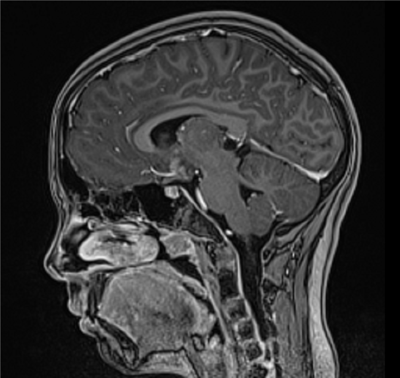
Figure 1 Suprasellar germinoma in a twelve-year-old girl.
The sagittal brain magnetic resonance imaging (MRI) scan demonstrates a suprasellar lesion with lobular contours, adjacent to the tuber cinereum and anterior commissure, involving the infundibulum of the pituitary gland and the optic chiasm. A and B: The lesion is isointense to the brain on non-contrast T1 weighted image (WI) (A) and shows homogenous contrast enhancement (B); C: The axial T2WI MRI scan shows the lesion isointense to the brain.
Figure 2 Brain magnetic resonance imaging after the end of chemotherapy.
Magnetic resonance imaging shows no residual tumor.
- Citation: Roganovic J, Saric L, Segulja S, Dordevic A, Radosevic M. Panhypopituitarism caused by a suprasellar germinoma: A case report. World J Clin Cases 2024; 12(10): 1844-1850
- URL: https://www.wjgnet.com/2307-8960/full/v12/i10/1844.htm
- DOI: https://dx.doi.org/10.12998/wjcc.v12.i10.1844


