Copyright
©The Author(s) 2024.
World J Clin Cases. Apr 6, 2024; 12(10): 1728-1732
Published online Apr 6, 2024. doi: 10.12998/wjcc.v12.i10.1728
Published online Apr 6, 2024. doi: 10.12998/wjcc.v12.i10.1728
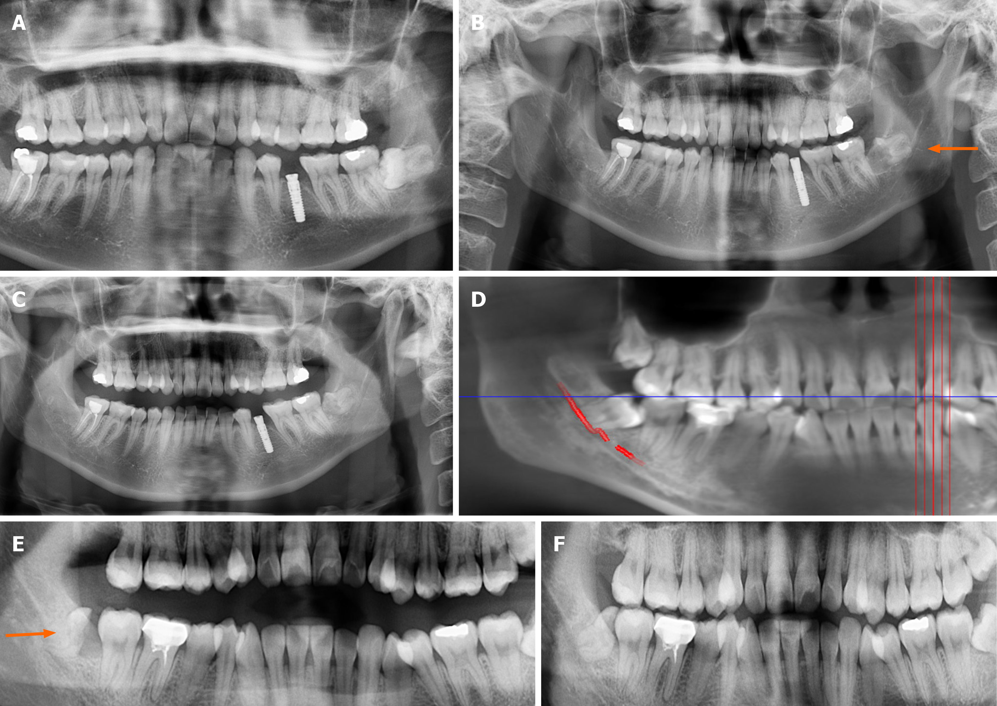
Figure 1 Panoramic views showing changes in relationship between the inferior alveolar nerve and impacted mandibular third molar root position by partial grinding in two cases (arrow indicates the mandibular canal).
A: Preoperative panoramic radiograph of Case 1 depicting direct contact between the inferior alveolar nerve (IAN) and the impacted mandibular third molar (IMM3); B: Post-partial grinding panoramic radiograph of Case 1, demonstrating sufficient eruption space between the impacted tooth and adjacent teeth; C: Six-month post-partial grinding view illustrating space between the root of the IMM3 and the superior wall of the IAN canal in Case 1; D: Preoperative panoramic radiograph of Case 2 revealing the IMM3 passing through the IAN; E: Post-partial removal of IMM3 (Phase 1) panoramic radiograph of Case 2, displaying adequate eruption space between the impacted tooth and adjacent teeth; F: Panoramic view after 6 mo of partial grinding indicating that in Case 2, the root of the IMM3 had moved away from the IAN canal following 6 months of partial grinding.
- Citation: Luo GM, Yao ZS, Huang WX, Zou LY, Yang Y. Two-stage extraction by partial grinding of impacted mandibular third molar in close proximity to the inferior alveolar nerve. World J Clin Cases 2024; 12(10): 1728-1732
- URL: https://www.wjgnet.com/2307-8960/full/v12/i10/1728.htm
- DOI: https://dx.doi.org/10.12998/wjcc.v12.i10.1728


