©The Author(s) 2024.
World J Clin Cases. Jan 6, 2024; 12(1): 142-147
Published online Jan 6, 2024. doi: 10.12998/wjcc.v12.i1.142
Published online Jan 6, 2024. doi: 10.12998/wjcc.v12.i1.142
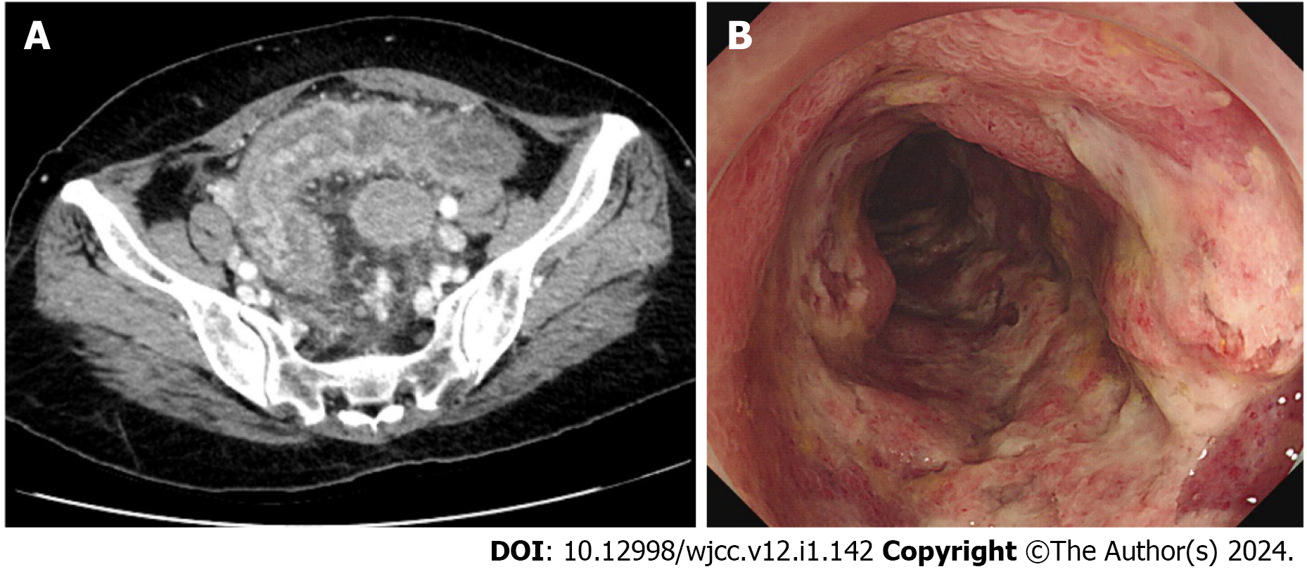
Figure 1 Pre-treatment imaging.
A: Abdominal computed tomography showed thickening of the sigmoid colon walls, pericolic fat infiltration, and a small amount of ascites; B: Multiple erosions and ulcers surrounding the area below the anastomosis site were observed during colonoscopy.
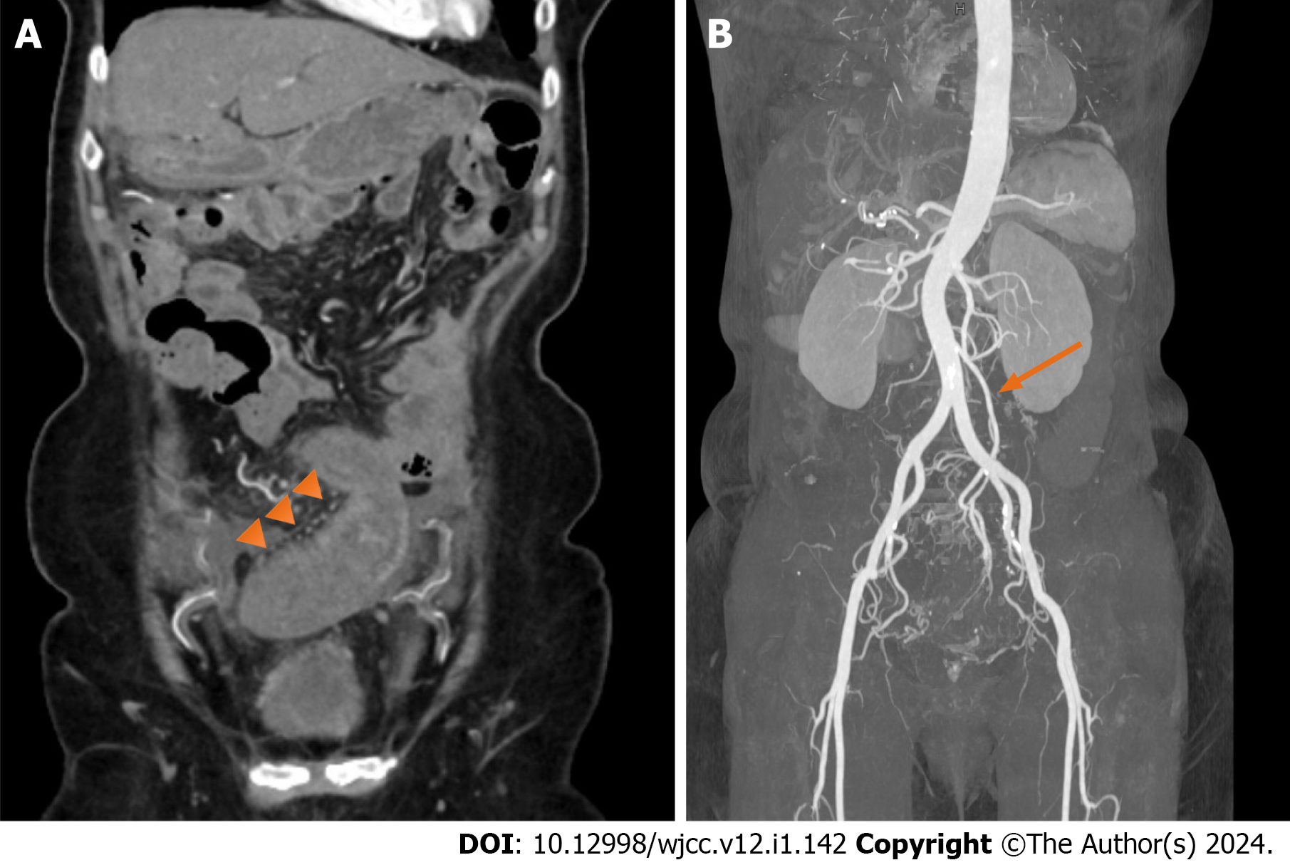
Figure 2 Pre-treatment computed tomography angiography.
A: Computed tomography angiography showed engorgement of the sigmoid vasa recta (triangular arrows); B: Blood flow in the inferior mesenteric, sigmoid colic, and superior rectal arteries remained preserved (arrow).
Figure 3 Post-treatment colonoscopy.
After 7 mo of treatment with anti-inflammatory therapy, colonoscopy showed no abnormal findings.
- Citation: Lee GW, Park SB. Congestive ischemic colitis successfully treated with anti-inflammatory therapy: A case report. World J Clin Cases 2024; 12(1): 142-147
- URL: https://www.wjgnet.com/2307-8960/full/v12/i1/142.htm
- DOI: https://dx.doi.org/10.12998/wjcc.v12.i1.142


