©The Author(s) 2024.
World J Clin Cases. Jan 6, 2024; 12(1): 130-135
Published online Jan 6, 2024. doi: 10.12998/wjcc.v12.i1.130
Published online Jan 6, 2024. doi: 10.12998/wjcc.v12.i1.130
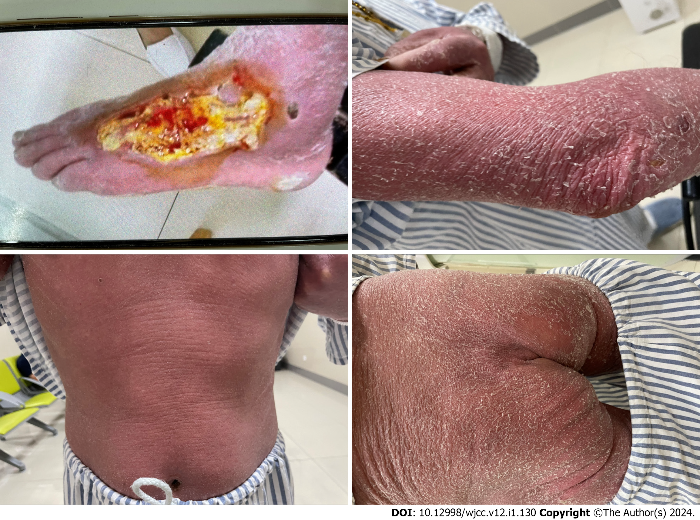
Figure 1 The clinical manifestations of patients.
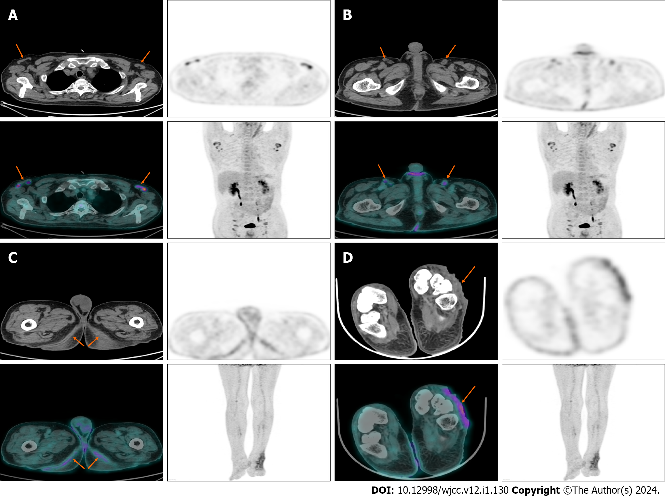
Figure 2 Show the positron emission tomography/computed tomography manifestations of some lesions.
A: Enlarged lymph nodes with increased fluoroDglucose (FDG) uptake in armpit; B: Enlarged lymph nodes with increased FDG uptake in groin; C: Thickening of both buttock skin with increased FDG uptake; D: Skin ulceration of left foot with increased FDG uptake.
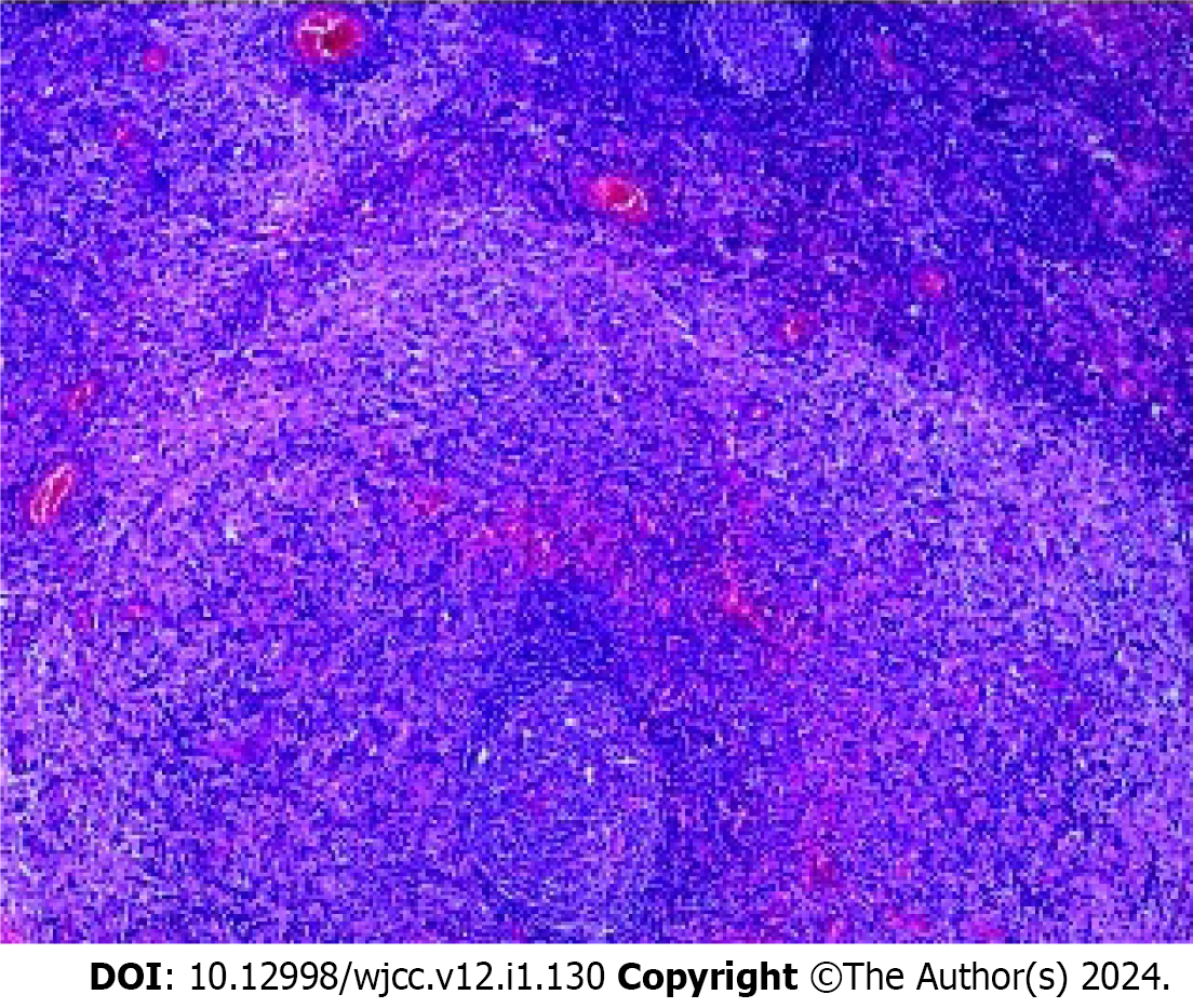
Figure 3 Abnormal T-cell proliferative lesions consistent with mycosis fungoides accumulative lymph nodes.
- Citation: Xu WB, Zhang YP, Zhou SP, Bai HY. Erythrodermic mycosis fungoides: A case report. World J Clin Cases 2024; 12(1): 130-135
- URL: https://www.wjgnet.com/2307-8960/full/v12/i1/130.htm
- DOI: https://dx.doi.org/10.12998/wjcc.v12.i1.130


