Copyright
©The Author(s) 2023.
World J Clin Cases. Mar 6, 2023; 11(7): 1650-1655
Published online Mar 6, 2023. doi: 10.12998/wjcc.v11.i7.1650
Published online Mar 6, 2023. doi: 10.12998/wjcc.v11.i7.1650
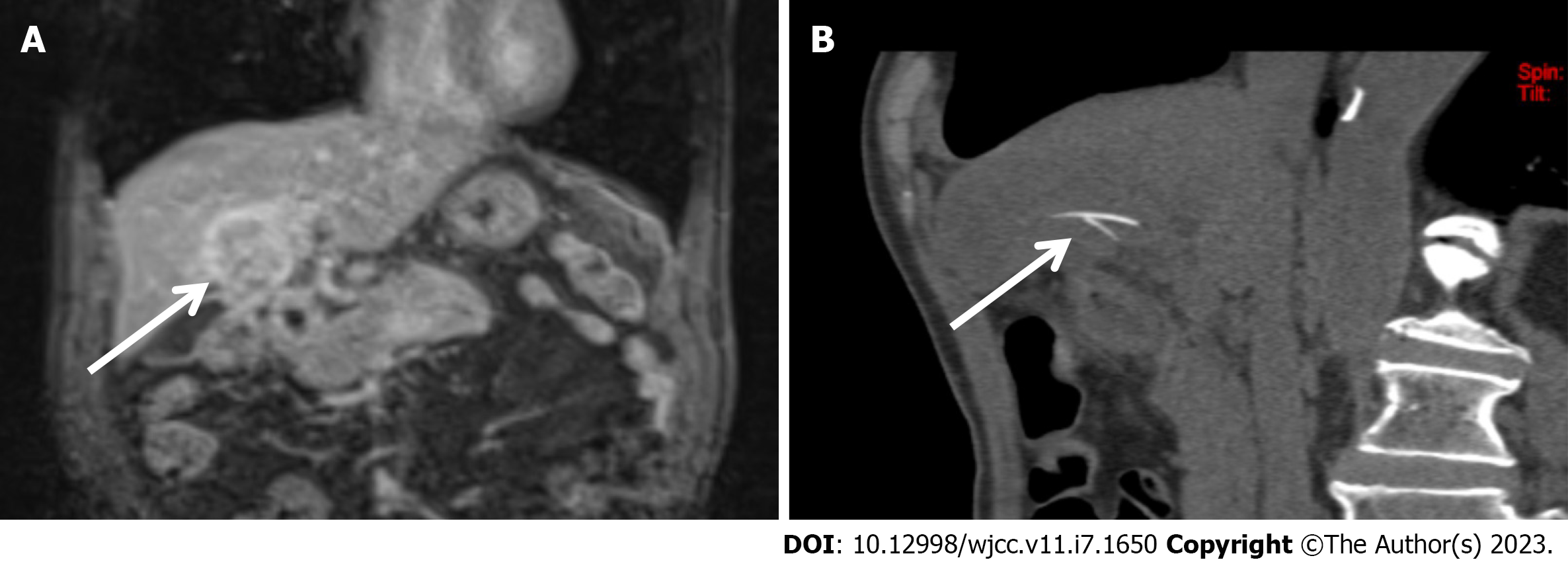
Figure 1 An 81-year-old man was admitted to our hospital with abdominal pain.
A: Magnetic resonance imaging showed mixed-signal shadows in the liver, and an enhanced scan showed uneven enhancement (arrow); B: Two months later, computed tomography images showed a strip-like shadow of bone density in the liver, and the edge of the shadow was sharp (arrow).
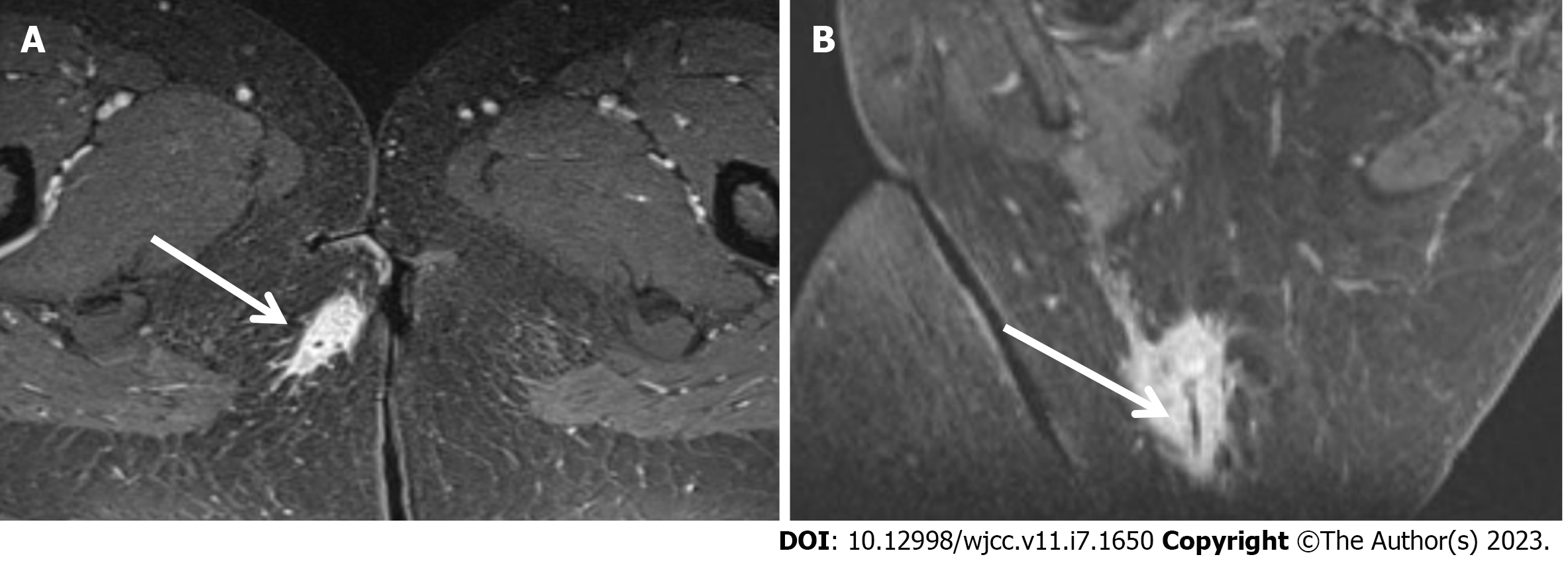
Figure 2 A 43-year-old woman was admitted to our hospital with hip pain.
A: Magnetic resonance imaging showed abnormal tubular signals behind the anal canal, obvious local enhancement on an enhanced scan, but no enhancement in some layers. Additionally, exudation was observed in the surrounding subcutaneous fat (arrow); B: A low signal bar with very clear and smooth edges was present, but sharp edges were evident on oblique coronal images (arrow).
- Citation: Ji D, Lu JD, Zhang ZG, Mao XP. Misdiagnosis of food-borne foreign bodies outside of the digestive tract on magnetic resonance imaging: Two case reports. World J Clin Cases 2023; 11(7): 1650-1655
- URL: https://www.wjgnet.com/2307-8960/full/v11/i7/1650.htm
- DOI: https://dx.doi.org/10.12998/wjcc.v11.i7.1650


