©The Author(s) 2023.
World J Clin Cases. Mar 6, 2023; 11(7): 1528-1548
Published online Mar 6, 2023. doi: 10.12998/wjcc.v11.i7.1528
Published online Mar 6, 2023. doi: 10.12998/wjcc.v11.i7.1528
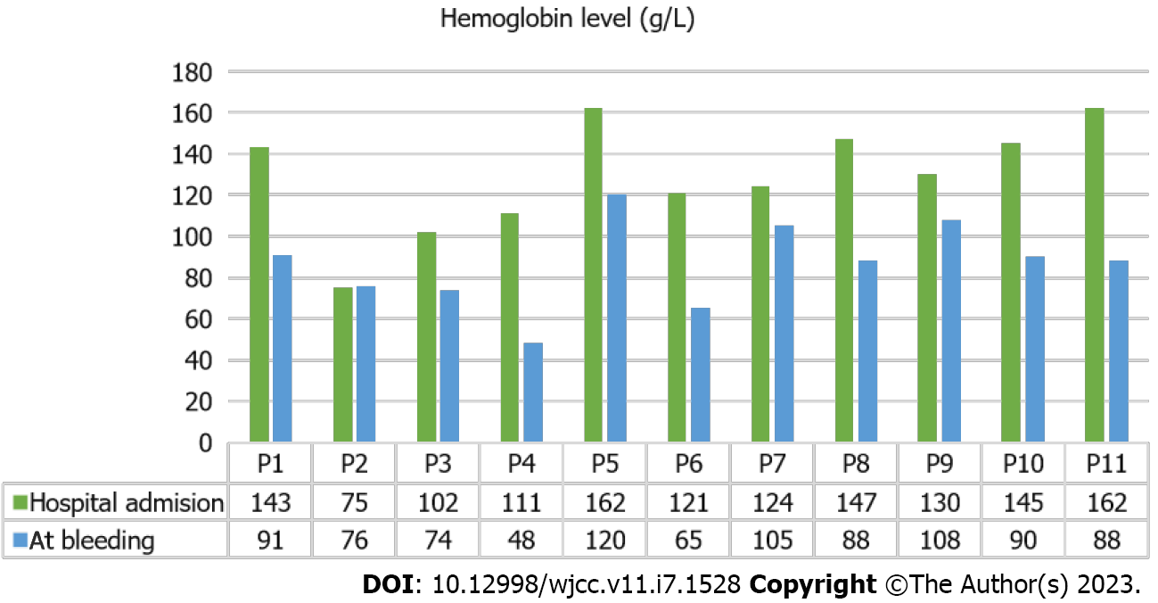
Figure 1 Dynamics of hemoglobin concentration in coronavirus disease 2019 patients.
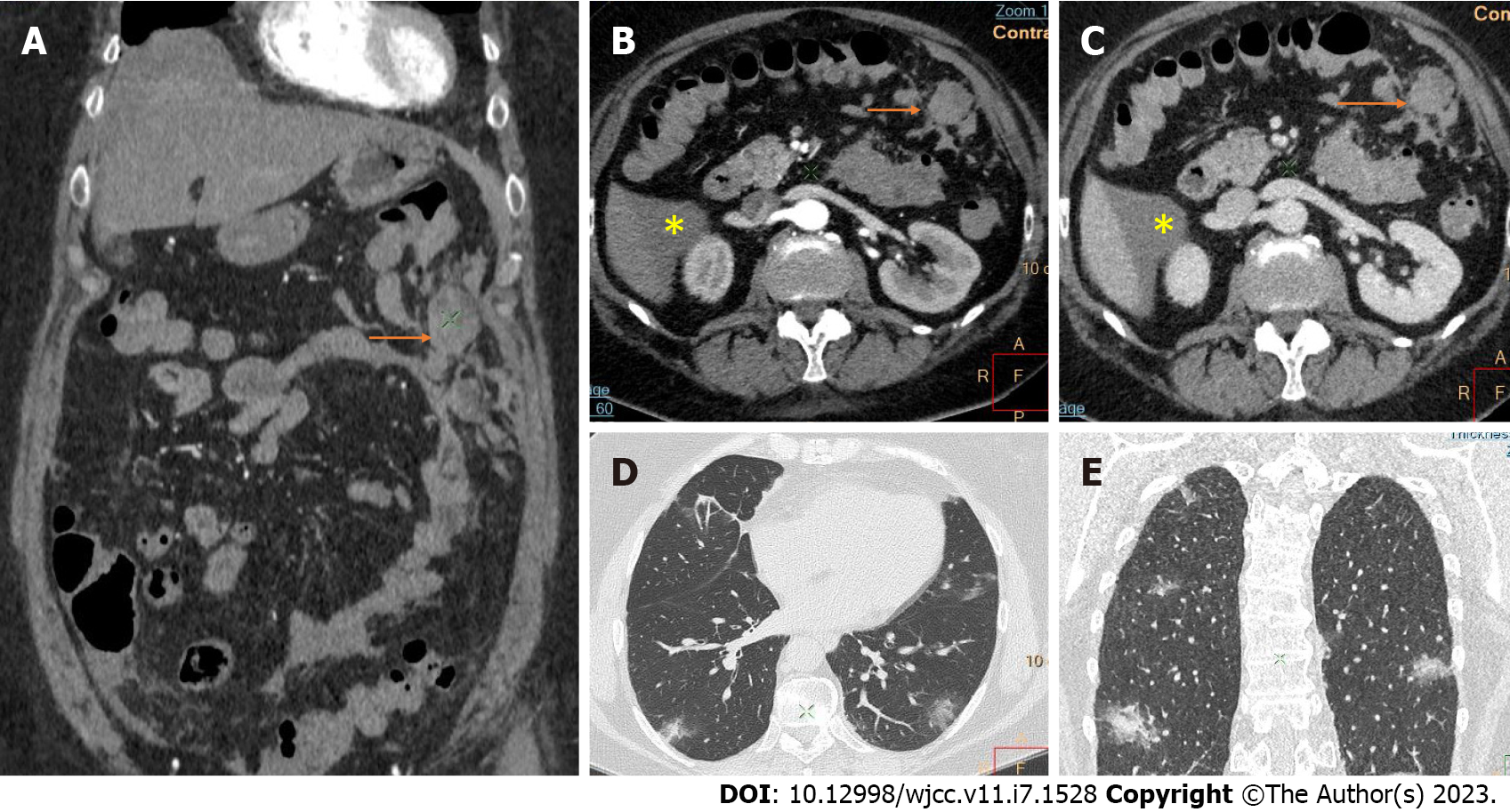
Figure 2 Computed tomography images of intraabdominal hemorrhage and lung involvement by coronavirus disease 2019 pneumonia.
A: Coronal reconstruction; B and C: Axial images with contrast demonstrate an intraabdominal hemorrhagic collection in the left upper quadrant without clear visualization of the site of bleeding (arrows) and small ascites close to the lower pole of the liver (asterisk); D and E: Bilateral lung "ground glass" consolidations in coronavirus disease 2019 pneumonia.
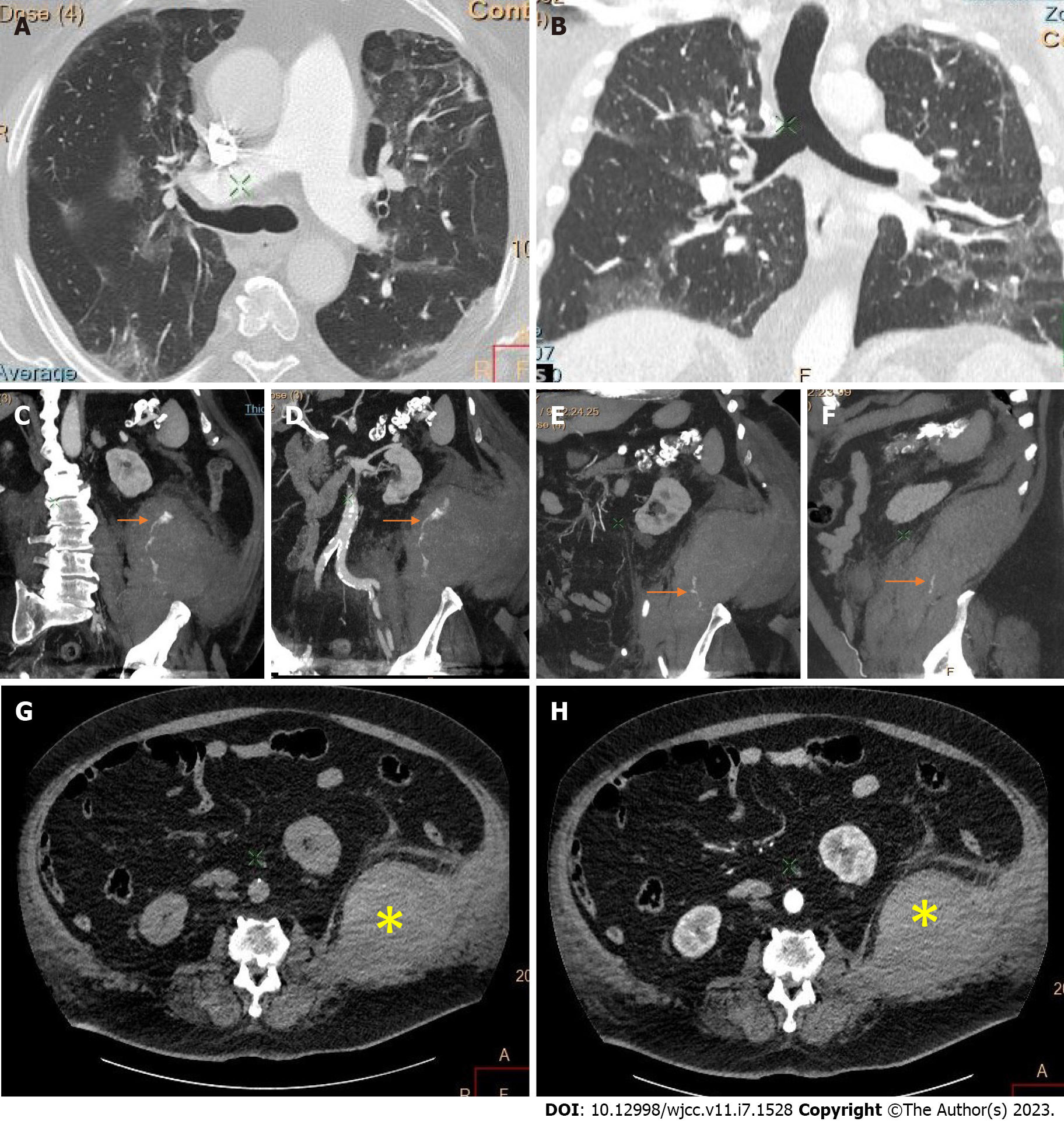
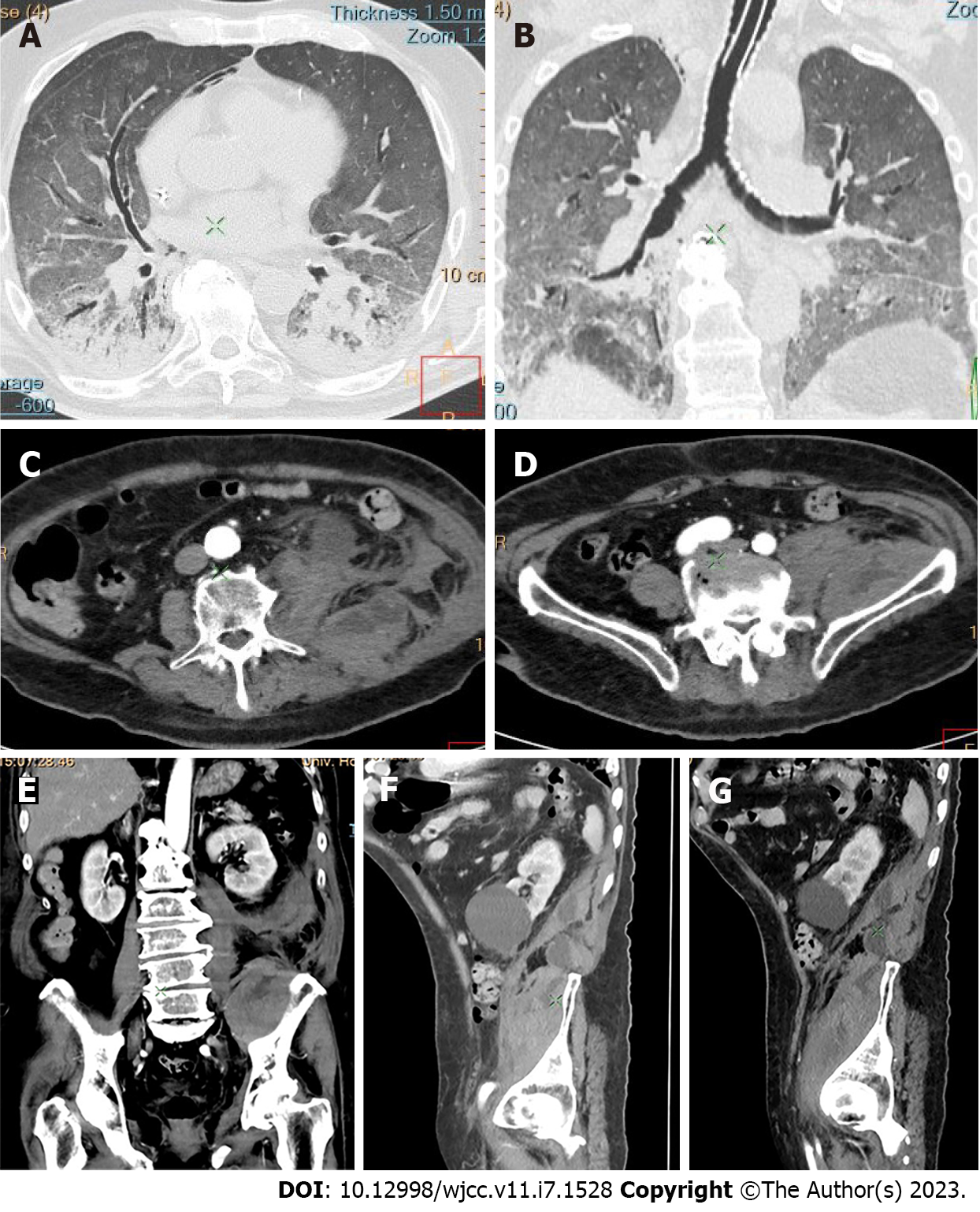
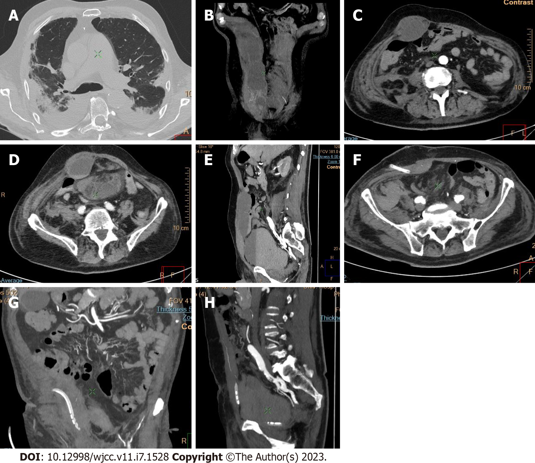
Figure 3 Thoracic and abdominal computed tomography of a large retroperitoneal hematoma.
A: Bilateral lung involvement with coronavirus disease 2019 (COVID-19) pneumonia in cross section; and B: In vertical section; C-F: Multiplanar reconstructions present the site of active bleeding and the jet from the extravasated contrast material (arrows); C-H: The hematoma arises from the internal, the external oblique, and the transverse abdominal muscles and invades the adjacent psoas, quadratus lumborum, and iliacus muscles on the left, which appear thickened (asterisk).
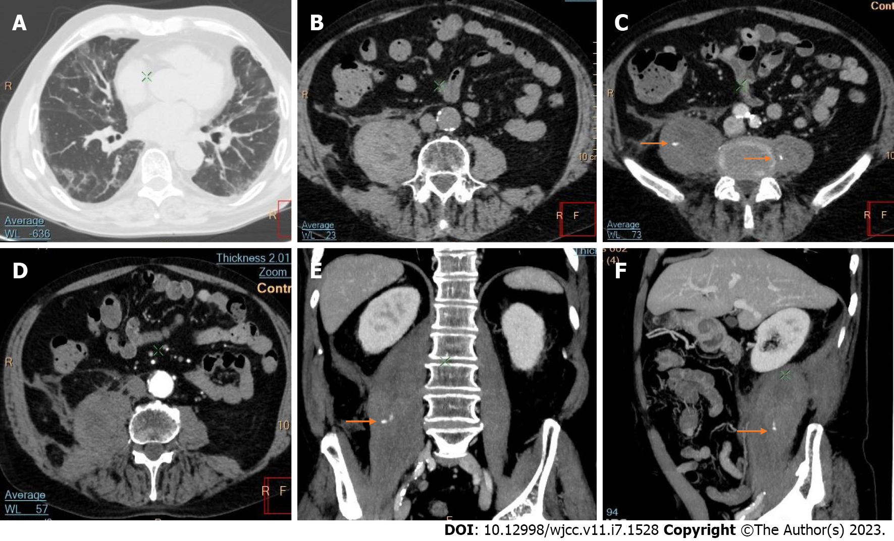
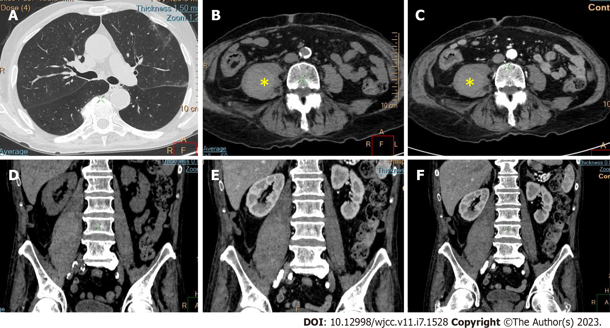
Figure 4 Axial computed tomography followed by coronal and sagittal reconstructions of retroperitoneal hematoma.
A: Axial computed tomography of inflammatory consolidations in bilateral coronavirus disease 2019 pneumonia; B-F: The hematoma originates from psoas major (B and C), the right quadratus lumborum (D), and iliacus muscles (E and F); C, E and F: Extravasated contrast material points to the site of active bleeding in the right psoas muscle (arrows).
Figure 5 Abdominal and pelvic computed tomography and dynamic contrast enhanced computed tomography of retroperitoneal hemorrhage on the left.
A: Bilateral lung consolidations with posterior predilection, predominantly involving the lower lobes, including “ground glass” opacities and vascular enlargement in cross section; B: Bilateral lung consolidations with posterior predilection, predominantly involving the lower lobes, including “ground glass” opacities and vascular enlargement in vertical section; C-E: Retroperitoneal hemorrhagic collection in the left flank and iliac fossa by postcontrast computed tomography axial and coronal images. Axial and coronal reformat images of dilatation of the psoas muscles; F and G: Sagittal reconstruction plane of the entire length of the iliac muscle affected by a hematoma.
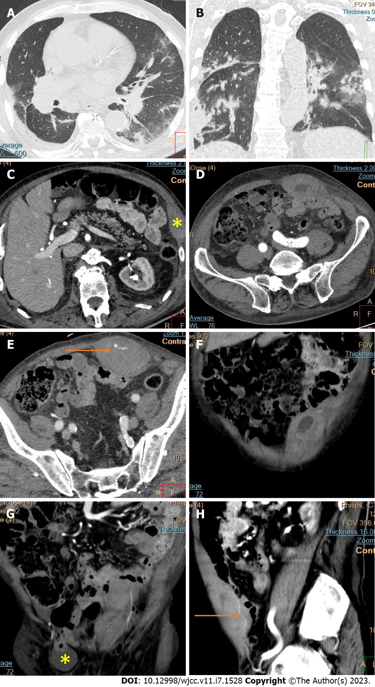
Figure 6 Computed tomography of active bleeding hematoma in the left rectus abdominis muscle.
A: Pulmonary “ground glass” opacification and interstitial thickening due to coronavirus disease 2019 (COVID-19) pneumonia in cross section; B: Pulmonary “ground glass” opacification and interstitial thickening due to COVID-19 pneumonia in vertical section; C: Hematoma between the internal and external abdominal muscles on the left (asterisk); D-H: The exact site of bleeding from the inferior epigastric artery on axial post-contrast images and coronal and sagittal maximum intensity projection reconstructions (arrows); G: Right-sided inguinal herniation containing a small bowel segment as an additional finding (asterisk).
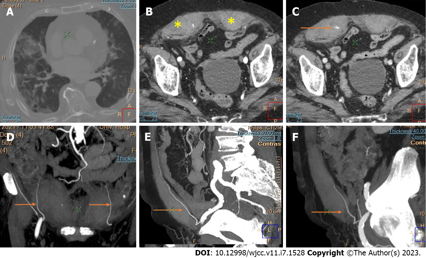
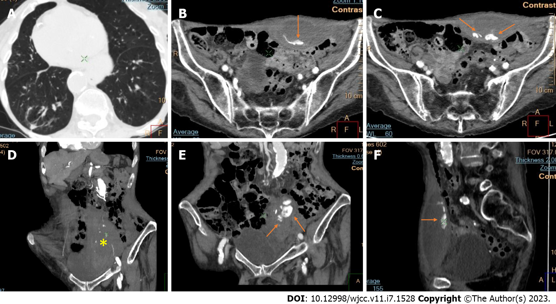
Figure 7 Axial computed tomography with maximum intensity projection reconstructions show bilateral hematomas of the right abdominal muscles.
A: Bilateral “ground glass” opacities in the context of coronavirus disease 2019 pneumonia; B and C: Post-contrast axial images; D-F: coronal and sagittal reconstructions demonstrate both hematomas (asterisks) and active bleeding from branches of the inferior epigastric artery (arrows).
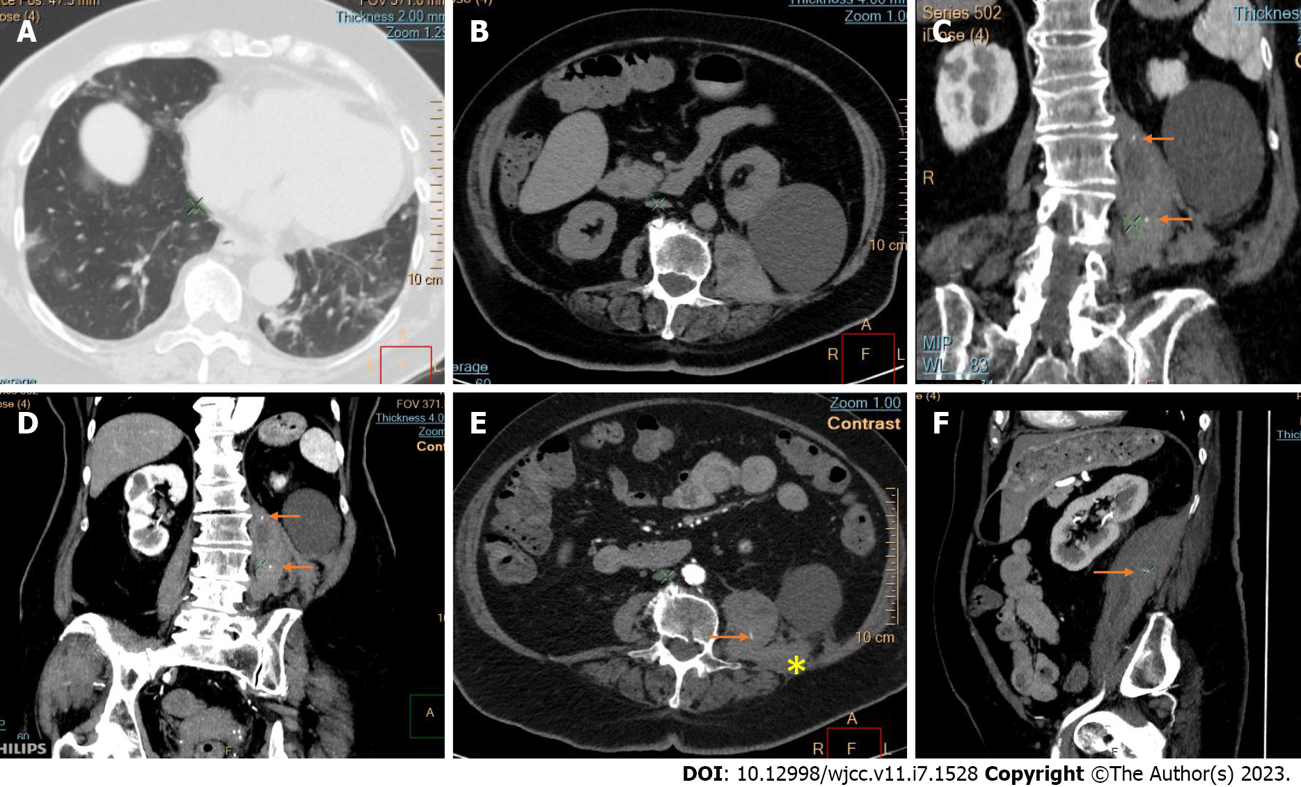
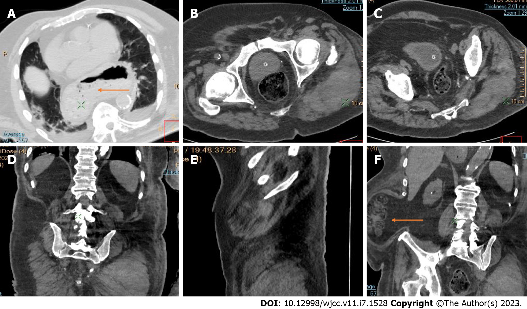
Figure 8 Abdominal pre- and post-contrast images and thoracic computed tomography in a patient with coronavirus disease 2019 and hematoma of the left psoas muscle.
A: “Crazy caving” pattern and linear interstitial thickening in the basal lung segments in organizing coronavirus disease 2019 pneumonia; B-F: A large hematoma of the left psoas muscle with infiltration of quadratus lumborum (asterisk); B-E: Large simple cortical cyst of the left kidney; C-F: Active bleeding from lumbar branches of the iliolumbar artery is indicated on post-contrast computed tomography images (arrows).
Figure 9 Large hematoma of the left gluteus maximus muscle in coronavirus disease 2019 pneumonia.
A: Lung parenchymal involvement by coronavirus disease 2019 pneumonia; B-F: Native computed tomography axial images and coronal and sagittal maximum intensity projection reconstructions visualize an unusual localization of anticoagulant-related muscular hematoma; F: Giant hiatal hernia and right lateral abdominal wall herniation (arrows) as additional findings.
Figure 10 Postcontrast computed tomography of a massive hematoma of the anterior abdominal wall.
A: This patient was recovering from coronavirus disease 2019 pneumonia with some residual post-inflammatory basal consolidations of the right lung; B-E: Depot of extravasated contrast is seen in the left rectus abdominis muscle (arrows); F: Clear bleeding from the left inferior epigastric artery (asterisk).
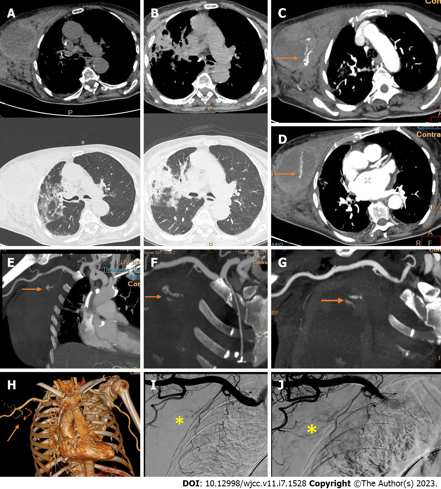
Figure 11 Computed tomography angiography and digital subtraction angiography of a giant right-sided chest wall hematoma in a patient with coronavirus disease 2019 pneumonia.
A-D: Hematoma arising from and infiltrating between the pectoral muscles, with “crazy paving” interstitial inflammatory changes in the right lung; C and D: Post-contrast axial images and coronal reconstructions with two pooling sites of extravasated contrast material within the hematoma (arrows); E-G: Computed tomography angiography maximum intensity projection images; H: Volume rendering three-dimensional reconstructions enable the visualization of a jet from the bleeding vessels (terminal branches of the superior and lateral thoracic artery (arrows). No bleeding from the pectoral branch of the thoracoacromial artery is evident; I and J: Digital subtraction angiograms confirm the predefined sites of bleeding without need of embolization due to bleeding reduction by the compression of those small vessels by the giant hematoma (asterisk).
Figure 12 Thoracic and pelvic computed tomography images of hematoma of the anterior abdominal wall.
A: Diffuse “ground glass” opacifications from coronavirus disease 2019 bilateral pneumonia; B and C: Pre-contrast axial and coronal images; D-F: Coronal, axial, and sagittal reconstructions with intravenous contrast show a mild hematoma in the left rectus abdominis muscle due to bleeding from the left inferior epigastric artery.
Figure 13 Axial multidetector computed tomography and maximum intensity projections in coronavirus disease 2019 pneumonia; right retroperitoneal and right abdominal wall hematoma.
A: Bilateral pleural effusions and reticular interstitial thickening in organized coronavirus disease 2019 pneumonia; B-E: Nodular thickening of the muscles of the anterior abdominal wall; C, D, F and G: Bloody infiltration of the right iliopsoas and iliacus muscles and posterior renal fascia with probable relationship to the right inferior epigastric artery; F-H: Pelvic drainage of re-bleeding during re-hospitalization.
Figure 14 Computed tomography images of right psoas major hematoma.
A: Minor peripheral and subpleural consolidations of the left lung in coronavirus disease 2019 pneumonia; B and C: Axial native and post-contrast images with enlargement of the right psoas muscle (asterisk); D-F: Dilated and hyperdense right psoas muscle by native coronal reconstructions and the same after contrast injection.
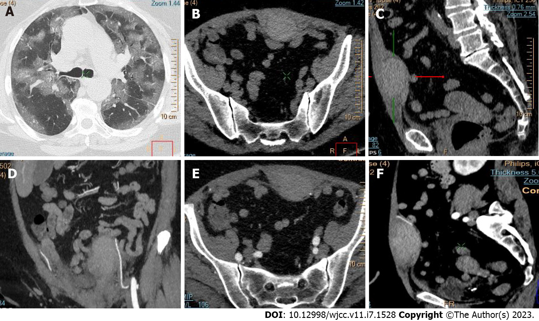
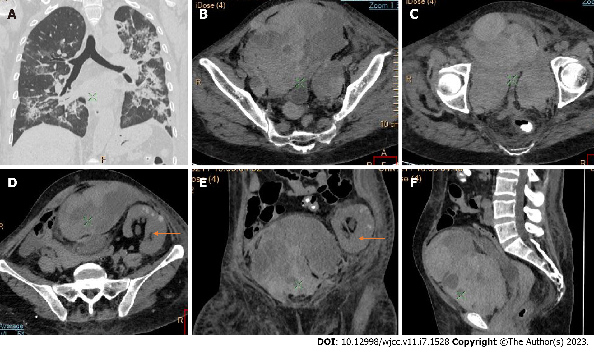
Figure 15 Computed tomography of enormous pre-peritoneal hematoma with intraabdominal and pelvic localization.
A: Severe bilateral consolidations with “crazy paving” due to coronavirus disease 2019 pneumonia; B-D: Axial images; E and F: Coronal reconstructions demonstrate a heterogeneous mass with mixed density; D and E: Transplanted kidney in the left iliac fossa (arrows).
- Citation: Evrev D, Sekulovski M, Gulinac M, Dobrev H, Velikova T, Hadjidekov G. Retroperitoneal and abdominal bleeding in anticoagulated COVID-19 hospitalized patients: Case series and brief literature review. World J Clin Cases 2023; 11(7): 1528-1548
- URL: https://www.wjgnet.com/2307-8960/full/v11/i7/1528.htm
- DOI: https://dx.doi.org/10.12998/wjcc.v11.i7.1528


