©The Author(s) 2023.
World J Clin Cases. Mar 6, 2023; 11(7): 1513-1520
Published online Mar 6, 2023. doi: 10.12998/wjcc.v11.i7.1513
Published online Mar 6, 2023. doi: 10.12998/wjcc.v11.i7.1513
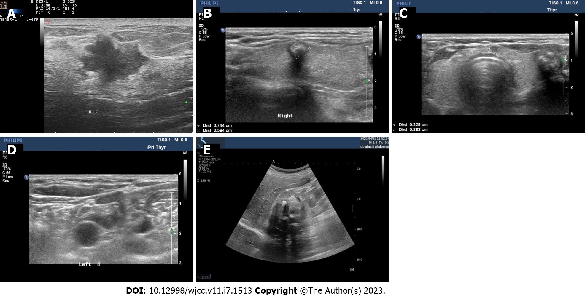
Figure 1 Ultrasound image.
A: Breast nodules; B: The right lobe nodule of the thyroid gland; C: Thyroid isthmus nodules; D: Nodules in the left lobe of the thyroid gland; E: The kidney nodules.
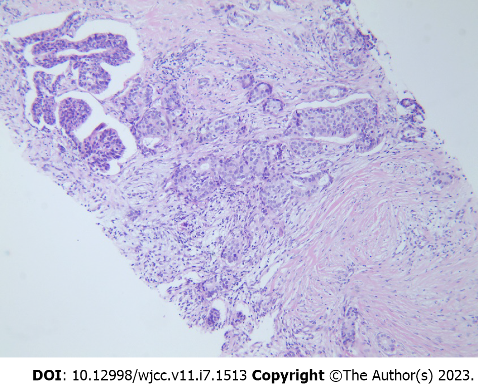
Figure 2 Puncture pathology of breast nodules.
Figure 3 Pathology.
A: Breast cancer; B: Kidney cancer; C: Thyroid cancer.
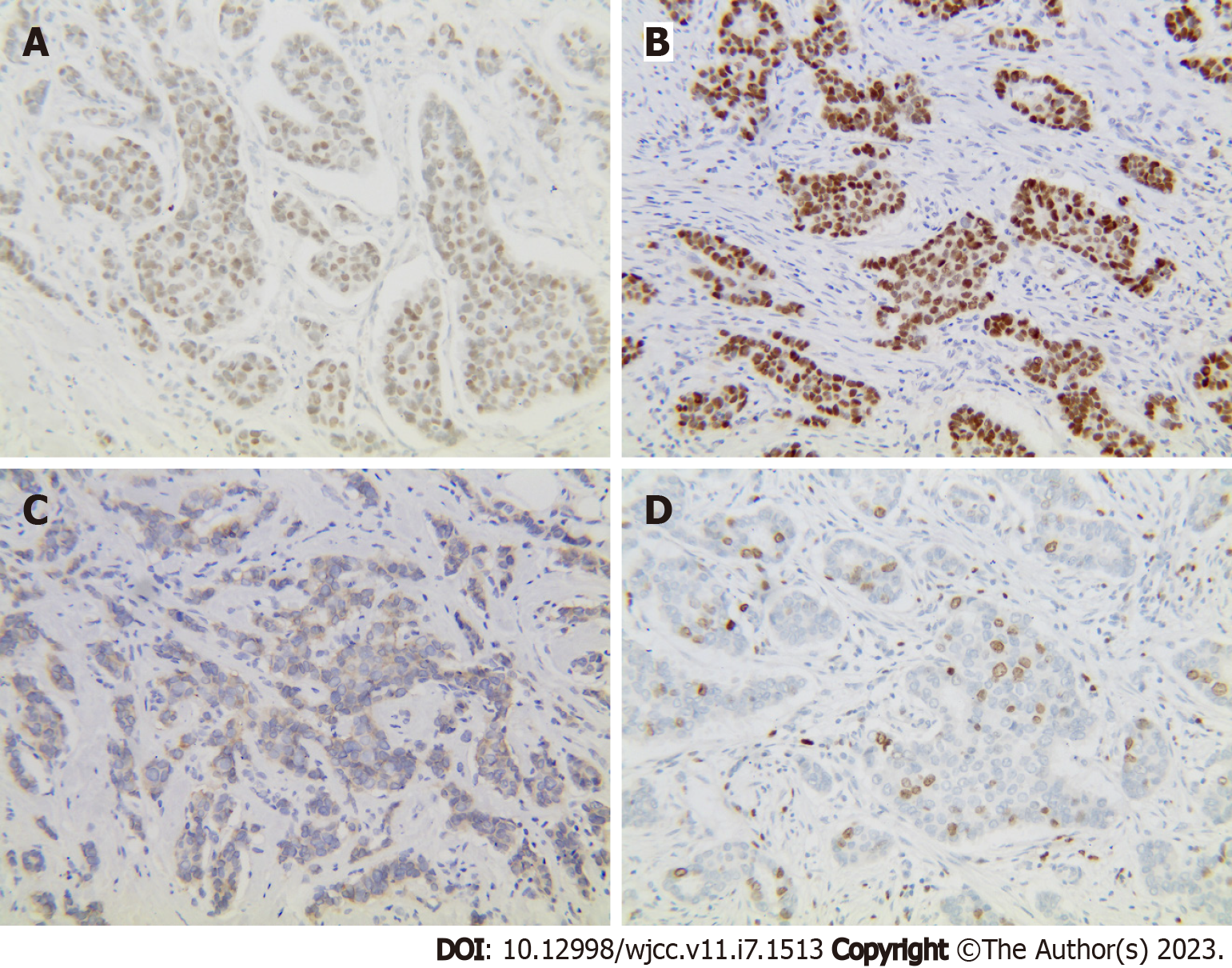
Figure 4 Immunohistochemical results.
A: Estrogen receptor (+, medium intensity, approximately 80%, 1604/2000 cells); B: Progesterone receptor (+, medium-strong intensity, approximately 80%, 1596/2000 cells); C: CerbB-2 (1+); D: Ki67 (approximately 30%+).
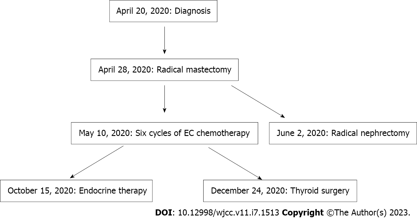
Figure 5 Timeline of the treatment.
EC: Endometrial cancer.
- Citation: Jia MM, Yang B, Ding C, Yao YR, Guo J, Yang HB. Synchronous multiple primary malignant neoplasms in breast, kidney, and bilateral thyroid: A case report. World J Clin Cases 2023; 11(7): 1513-1520
- URL: https://www.wjgnet.com/2307-8960/full/v11/i7/1513.htm
- DOI: https://dx.doi.org/10.12998/wjcc.v11.i7.1513


