Copyright
©The Author(s) 2023.
World J Clin Cases. Feb 26, 2023; 11(6): 1356-1364
Published online Feb 26, 2023. doi: 10.12998/wjcc.v11.i6.1356
Published online Feb 26, 2023. doi: 10.12998/wjcc.v11.i6.1356
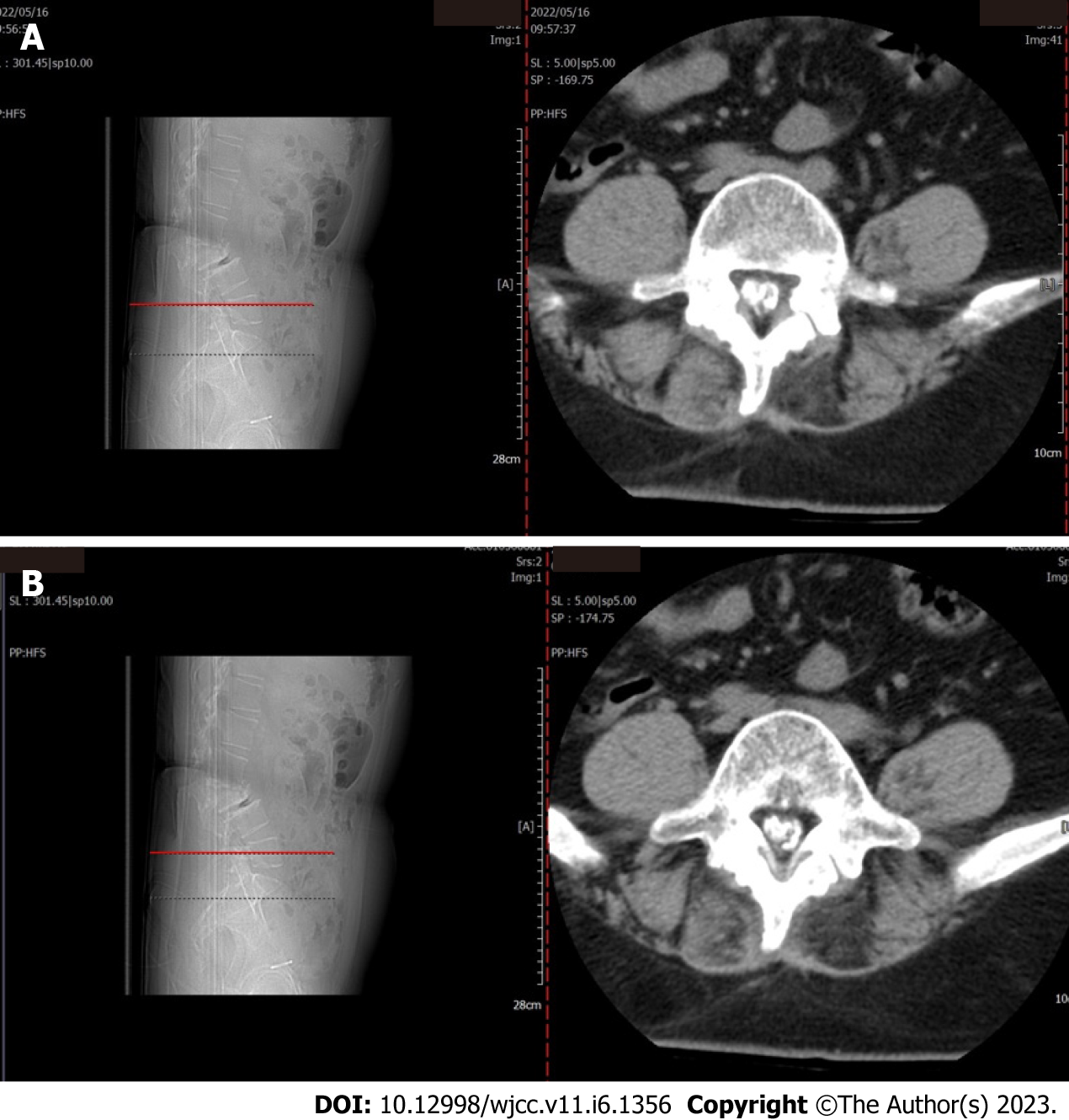
Figure 1 Lumbar spine computed tomography.
L5 plane end filament and cauda equina nerve calcification signs. A: L5 upper edge of vertebral body; B: L5 center of vertebral body.
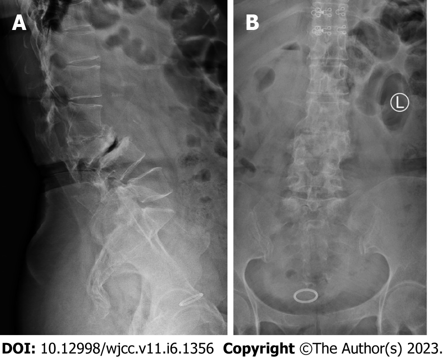
Figure 2 The X-ray film of lumbar vertebrae.
The sequence of lumbar vertebrae is abnormal, L3 vertebral body is diseased and shifted backward, L2-3 intervertebral space disappears, L2-3, L3-4, L4-5, L5-S1 intervertebral space narrows, and all vertebrae and vertebral facet joints have hyperosteogeny. A: Orthostatic lumbar X-ray film; B: Lateral lumbar X-ray film.
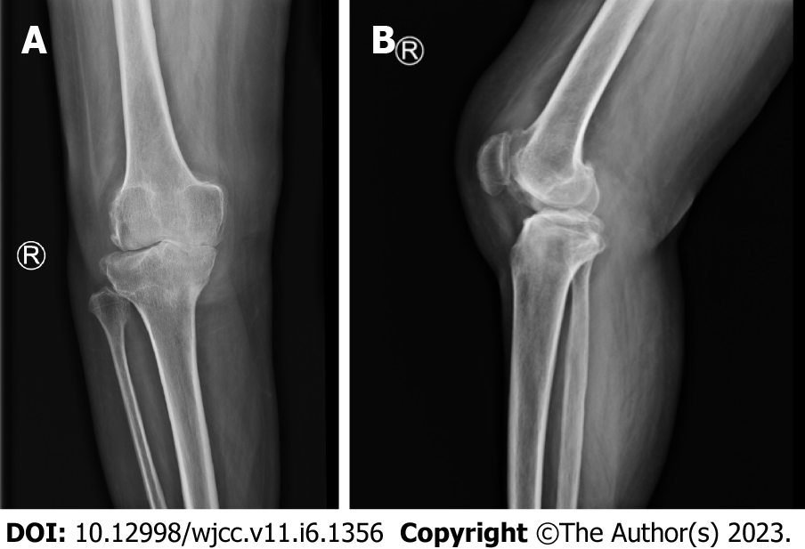
Figure 3 The X-ray film of the right knee joint.
The right knee joint space was narrow inside and wide outside, the articular surface was sclerotic, and the lower edge of the articular surface was spotted with low density. Hyperosteogeny appeared on the upper edge of patella, tibial plateau, tibial intercondylar crest and femoral condyle. The density of the suprapatellar bursa is increased and the surrounding soft tissue is swollen. A: Orthostatic Knee joint X-ray film; B: Lateral Knee joint X-ray film.
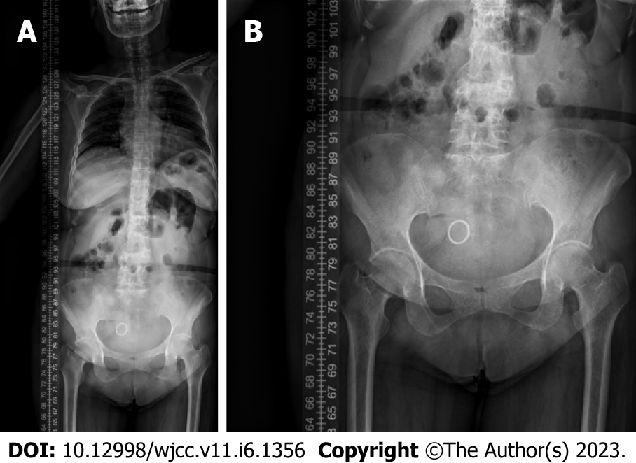
Figure 4 The X-ray film of the full length of the spine.
L3-4 vertebral body is diseased and shifted backward, the lumbar spine protrudes backward at the L2-3 plane, the intervertebral space at the L2-3 segment is narrowed, and the L3 vertebral body is wedge-shaped and flattened. A: The Orthostatic X-ray film of the full length of the spine; B: The Orthostatic X-ray film of lumbar spine and pelvis.
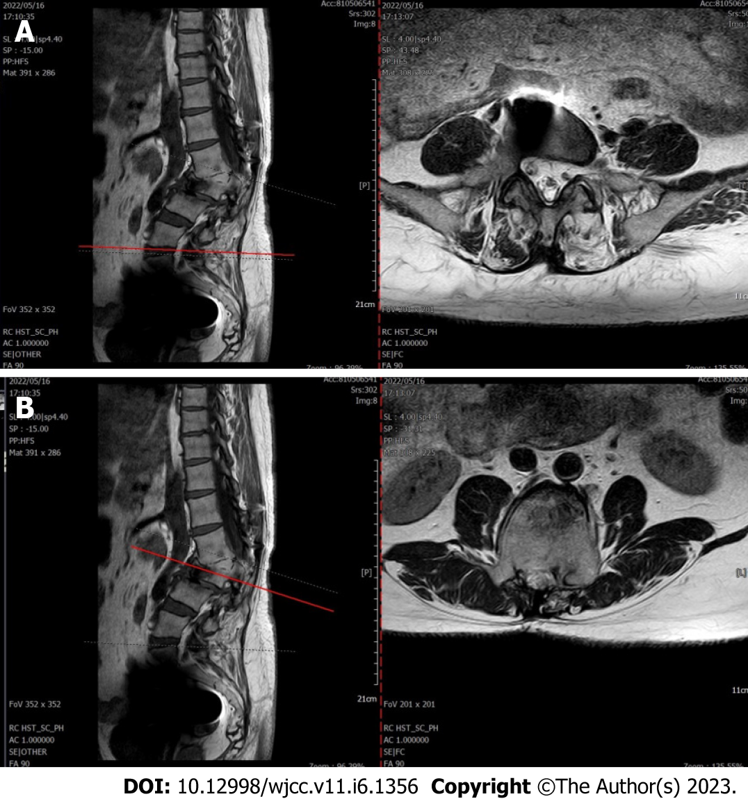
Figure 5 The lumbar spine magnetic resonance imaging.
Lumbar vertebrae protrude backward, L3 vertebrae show wedge-shaped changes, spinal canal stenosis on the same level of L3, intervertebral space narrowing on the same level of L2-3, endplate inflammation in the L3-4 intervertebral space, Schmorl node formation near T10-11 vertebral body, degeneration and bulge of L3-4, L4-5 and L5-S1 intervertebral discs, and many abnormal strip signals in the filum terminale. A: L5-S1 Plain magnetic resonance imaging (MRI) scan of L5-S1 intervertebral disc; B: L3 MRI plain scan.
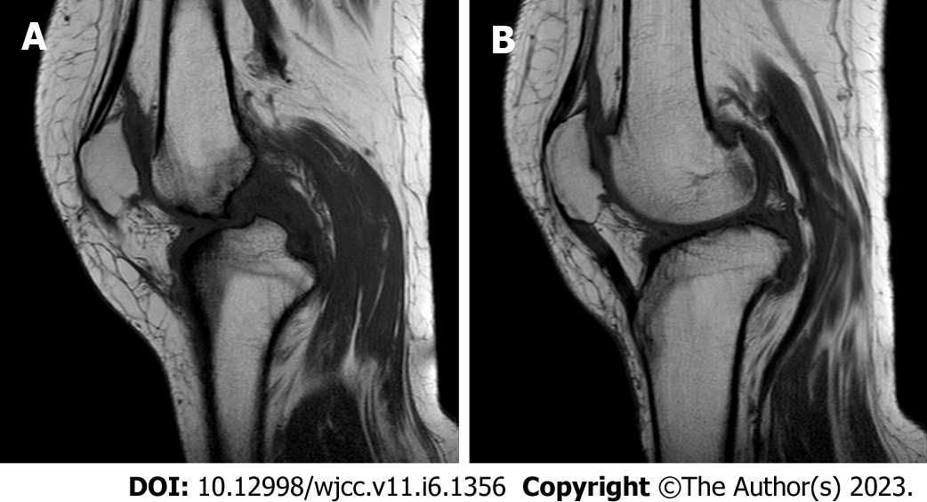
Figure 6 The right knee magnetic resonance imaging.
The cartilage of the medial femoral condyle and tibial plateau is worn, the cartilage is denatured, the posterior cruciate ligament of the right knee is torn, the medial collateral ligament is injured, the anterior and posterior corners of the medial and lateral meniscus of the right knee are worn, the right knee joint has effusion, and the medial head bursa of the gastrocnemius has effusion. A: Lateral magnetic resonance imaging (MRI) of knee; B: Oblique MRI of knee.
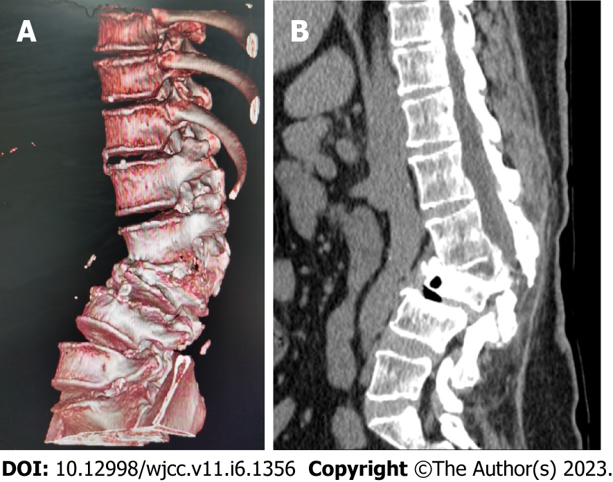
Figure 7 Lumbar vertebral body (plain scan + 3D reconstruction) computed tomography.
Lumbar vertebral body (plain scan + 3D reconstruction) computed tomography (CT) showed: L2-3 plane lumbar lordosis, L2-3 vertebral space narrowing, L3 vertebral body wedge-shaped flattening, L3, 4 vertebral body adjacent edge patchy high density. There are some bone defects in the lamina and spinous process of L3 and L4 vertebrae, and the vertebral canal at the same plane is deformed. A: Lumbar vertebral body CT (3D reconstruction); B: Lumbar vertebral body CT (plain scan).
- Citation: Liu YD, Deng Q, Li JJ, Yang HY, Han XF, Zhang KD, Peng RD, Xiang QQ. Post-traumatic cauda equina nerve calcification: A case report. World J Clin Cases 2023; 11(6): 1356-1364
- URL: https://www.wjgnet.com/2307-8960/full/v11/i6/1356.htm
- DOI: https://dx.doi.org/10.12998/wjcc.v11.i6.1356


