Copyright
©The Author(s) 2023.
World J Clin Cases. Feb 26, 2023; 11(6): 1318-1329
Published online Feb 26, 2023. doi: 10.12998/wjcc.v11.i6.1318
Published online Feb 26, 2023. doi: 10.12998/wjcc.v11.i6.1318
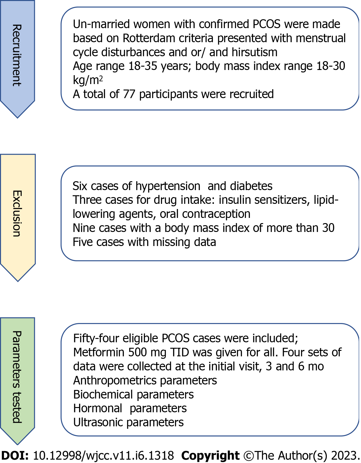
Figure 1 Study flowchart.
PCOS: Polycystic ovarian syndrome.
Figure 2 A sample of internal carotid artery Doppler.
A: It shows a real-time internal carotid artery Doppler; we have simulated the way of measuring the area under the curve by Paint app to the last wave. Note how different heights of the systolic and diastolic blood flow (demarcated with orange dots ), which are used to measure the area under the curve (demarcated as orange longitudinal lines); B: It shows the graphical simulation of the internal carotid artery Doppler wave with the different heights (demarcated in red dots) used to measure the systolic and diastolic blood flow for the calculation of the area under the curve.
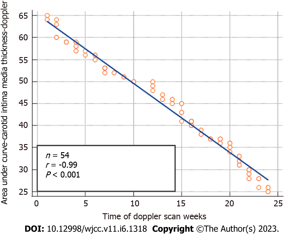
Figure 3 Highlights an inverse correlation of area under the curve of the internal carotid artery vs metformin treatment time (r = -0.
99, P < 0.001).
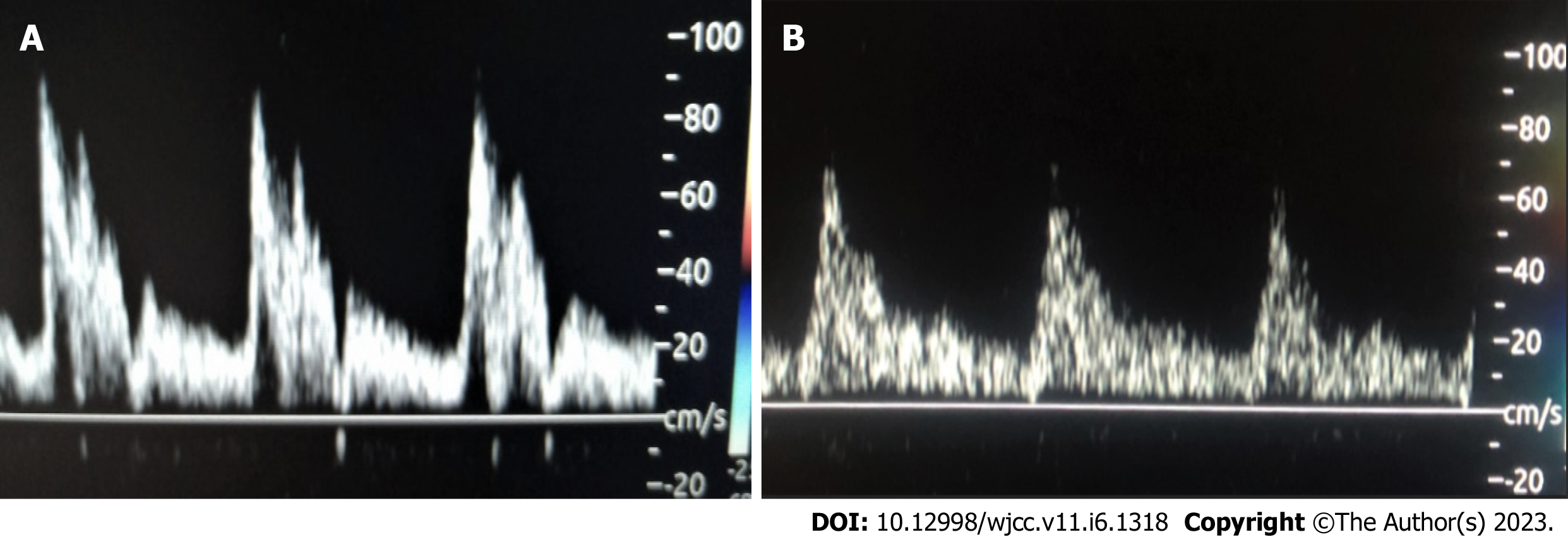
Figure 4 A real-time Doppler scan.
A: A real-time Doppler scan showing progressive reduction of the area under the curve of the internal carotid artery with treatment. Note the decrease in the peak of the wave in a subsequent visit 3 mo apart; from 80 to 60; B: A real-time Doppler scan showing the progressive reduction of the area under the curve of the internal carotid artery with treatment. Note the reduced wave peak to 60 following treatment compared to previous Figure 4A.
- Citation: Akram W, Nori W, Abdul Ghani Zghair M. Metformin effect on internal carotid artery blood flow assessed by area under the curve of carotid artery Doppler in women with polycystic ovarian syndrome. World J Clin Cases 2023; 11(6): 1318-1329
- URL: https://www.wjgnet.com/2307-8960/full/v11/i6/1318.htm
- DOI: https://dx.doi.org/10.12998/wjcc.v11.i6.1318


