©The Author(s) 2023.
World J Clin Cases. Dec 16, 2023; 11(35): 8404-8410
Published online Dec 16, 2023. doi: 10.12998/wjcc.v11.i35.8404
Published online Dec 16, 2023. doi: 10.12998/wjcc.v11.i35.8404
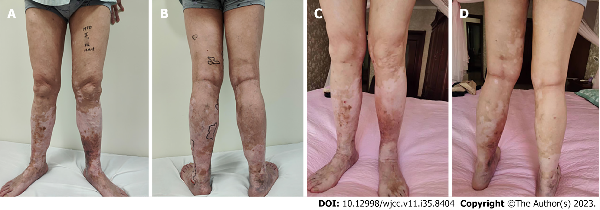
Figure 1 Comparison before and after surgery.
A and B: Before surgery, varicose veins and venous skin ulcers are observed on the front (A) and back (B) of both legs before surgery; C and D: 6 mo after surgery, the skin infection and ulcers have been treated and have disappeared on both the front (A) and back (B) of both legs.
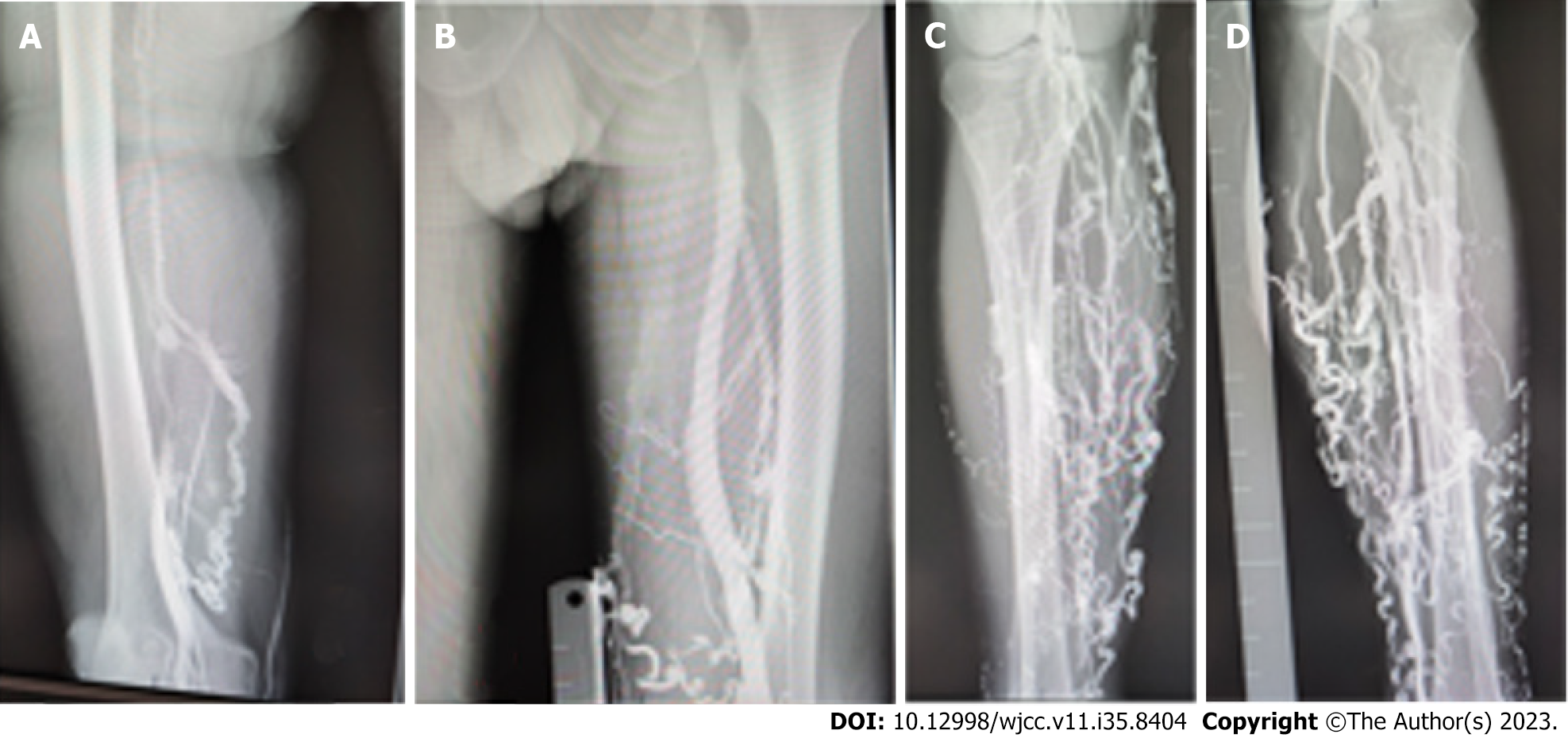
Figure 2 Venous angiography.
A and C: Multiple tortuous varicose veins are evident in the right leg. Venous thrombosis is suspected in the right thigh; B and D: The left leg also shows similar varicose veins with venous reflux extending to the knee.
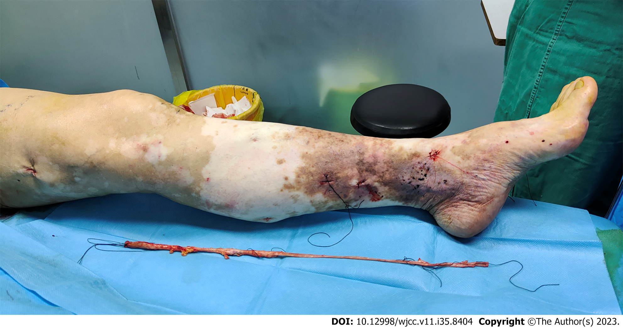
Figure 3 Varicose veins, edema, pigmentation, and vitiligo are observed on the lower extremities of the patient.
The ossified varicose vein has been surgically removed.
Figure 4 Dissected varicose vein shows thickening and hardening of the venous wall, with thrombus inside the vein and sparse erosion, ulceration, and blood clots on the intima of the vein.
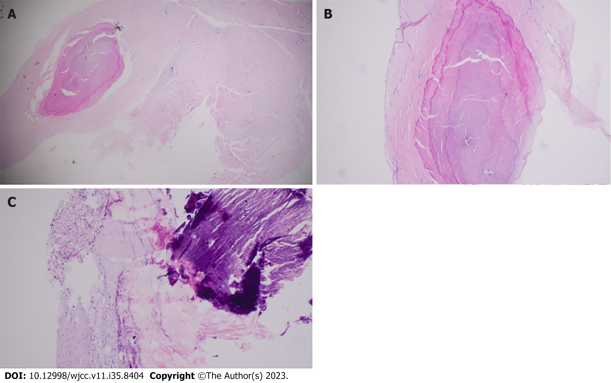
Figure 5 Phlebosclerosis: pathological examination of the vein resembling a wooden rod.
A: The cross-section of the vein during surgery shows venous fibrosis, calcification, and thickening of the vein wall. Extensive collagen deposition is observed on the vein wall, with hyaline degeneration and venous sclerosis causing closure of the venous lumen (× 40 magnification); B: × 100 magnification; C: Calcification in the venous lumen and inflammatory cell infiltration in the venous wall (× 100 magnification).
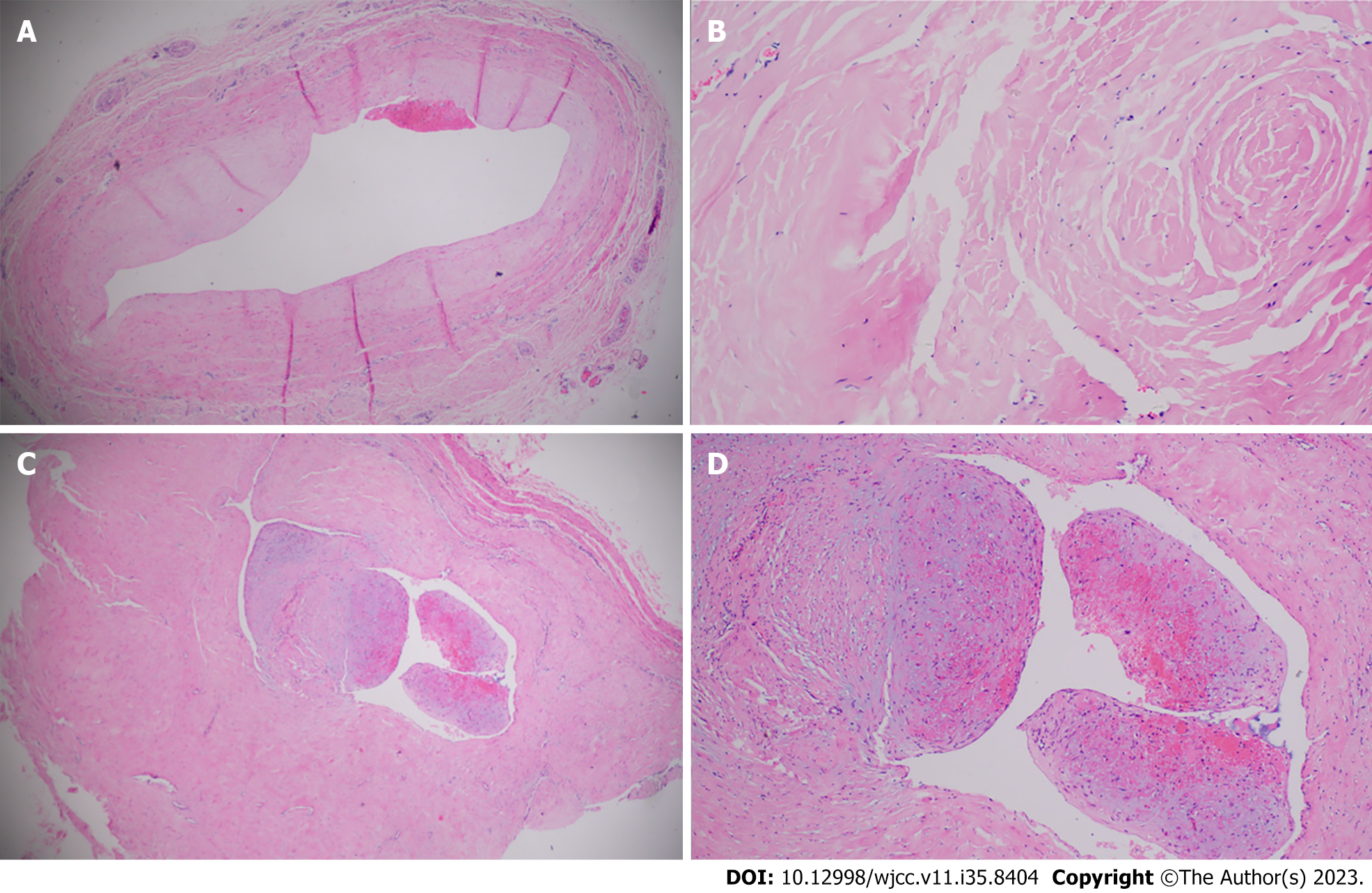
Figure 6 Phlebosclerosis: smooth muscle hyperplasia and granulation tissue.
A: Venous wall thickening and evident endothelial hyperplasia are observed. Thrombus adheres to the luminal endothelium, and the lumen is narrowed to varying degrees. (× 100 magnification); B: Smooth muscle hyperplasia is evident (× 200 magnification); C: Thrombus mechanization at the lumen and granulation tissue formation (× 40 magnification); D: Resultant luminal stenosis or occlusion (× 100 magnification).
- Citation: Ren SY, Qian SY, Gao RD. Phlebosclerosis: An overlooked complication of varicose veins that affects clinical outcome: A case report. World J Clin Cases 2023; 11(35): 8404-8410
- URL: https://www.wjgnet.com/2307-8960/full/v11/i35/8404.htm
- DOI: https://dx.doi.org/10.12998/wjcc.v11.i35.8404


