©The Author(s) 2023.
World J Clin Cases. Dec 16, 2023; 11(35): 8392-8398
Published online Dec 16, 2023. doi: 10.12998/wjcc.v11.i35.8392
Published online Dec 16, 2023. doi: 10.12998/wjcc.v11.i35.8392
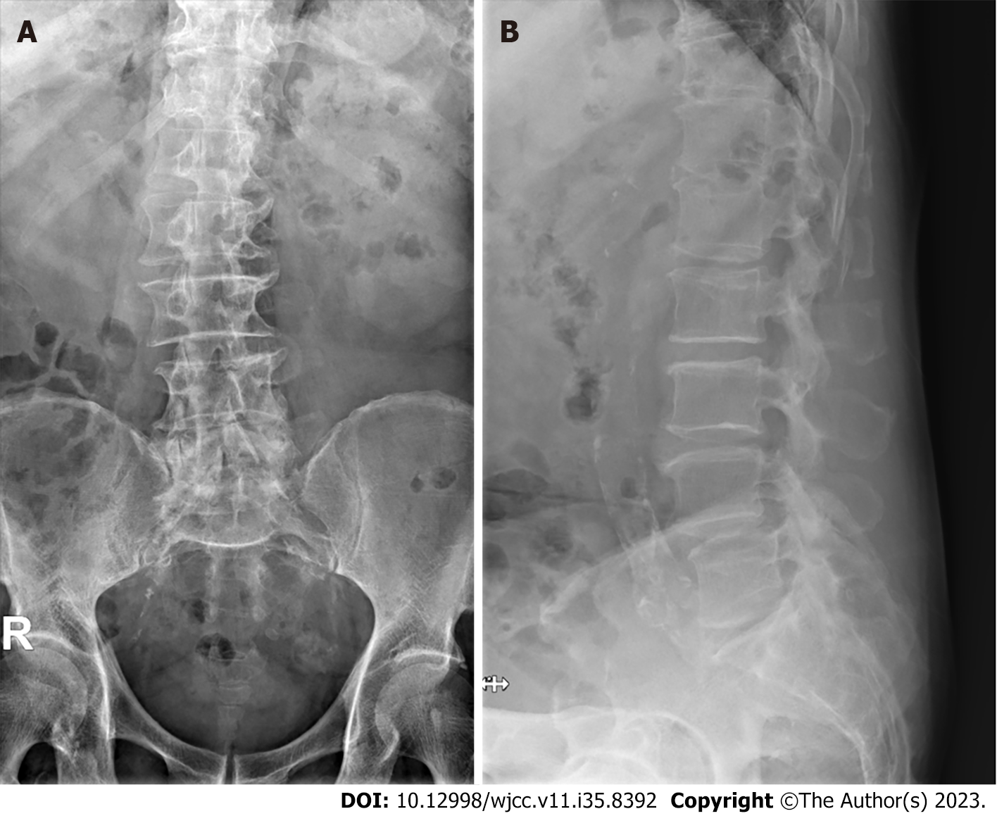
Figure 1 Plain radiographs of the lumbar spine.
A: Lumbar spine anteroposterior X-ray; B: Lumbar spine lateral flexion X-ray showing degenerative changes and spondylolisthesis L4 on L5.
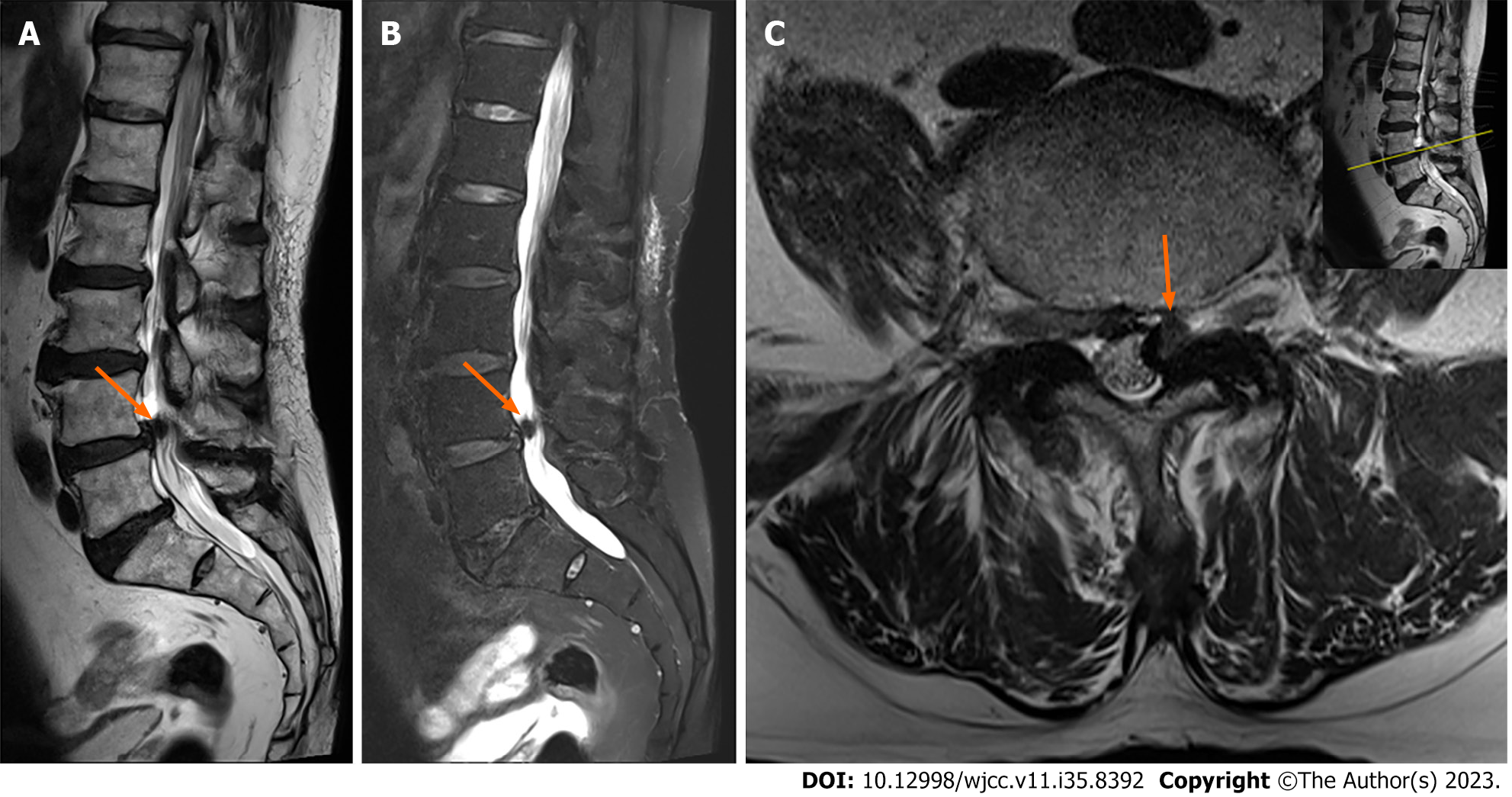
Figure 2 Preoperative magnetic resonance imaging of the lumbar spine.
A: Sagittal T2-weighted magnetic resonance imaging (MRI) showing a mass lesion with a hypointense signal at the L4-5 level (arrow); B: Sagittal fat-suppressed T2-weighted MRI showing a mass lesion with a hypointense signal at the L4-5 level (arrow); C: Axial T2-weighted MRI of the corresponding section showing left side dural sac compression by a mass (arrow).
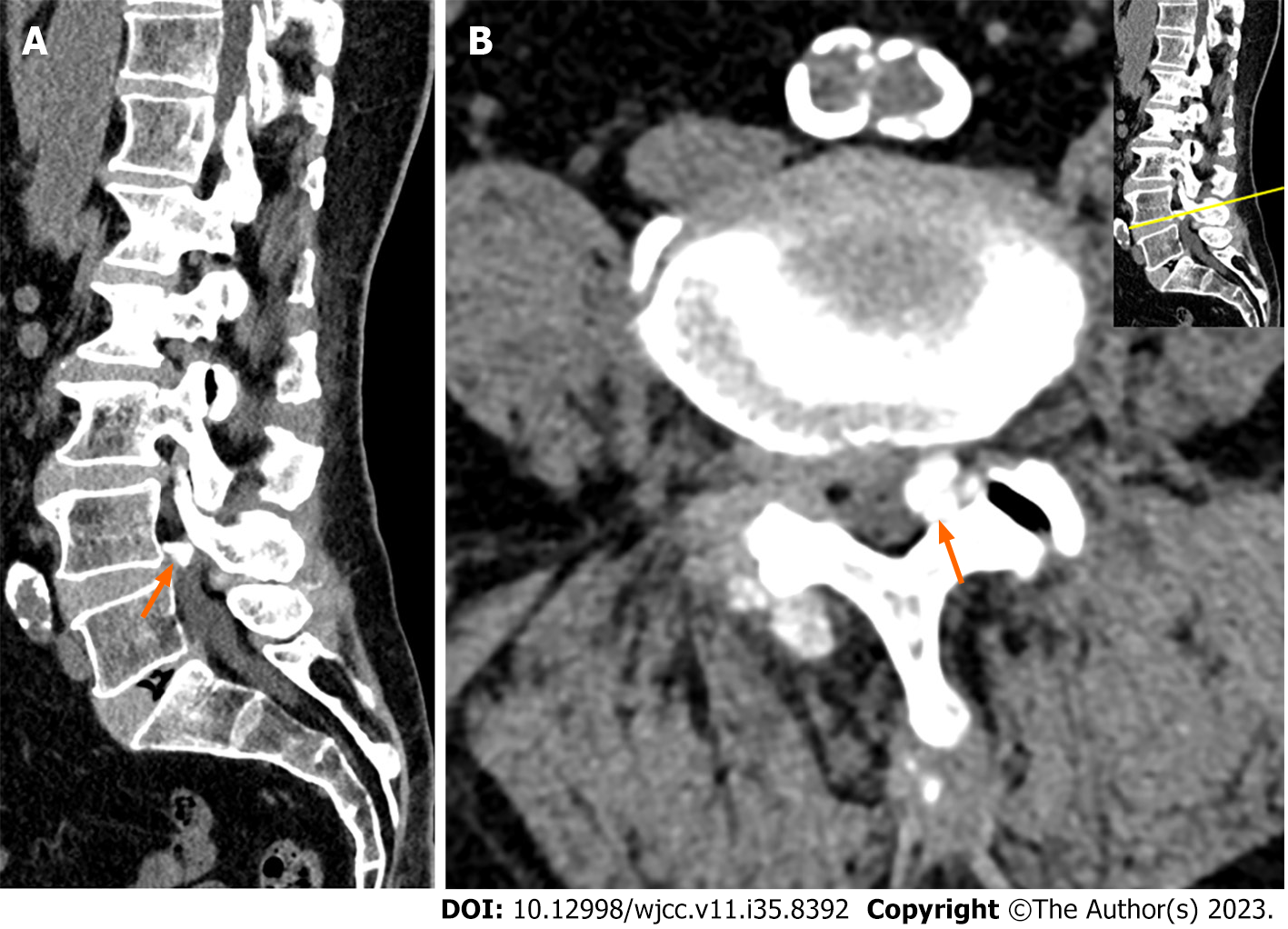
Figure 3 Computed tomography scans of the lumbar spine.
A: Sagittal computed tomography (CT) scan showing a calcified mass at the L4-5 level (arrow); B: Axial CT scan showing a calcified mass at the left side of the L4-5 level (arrow).
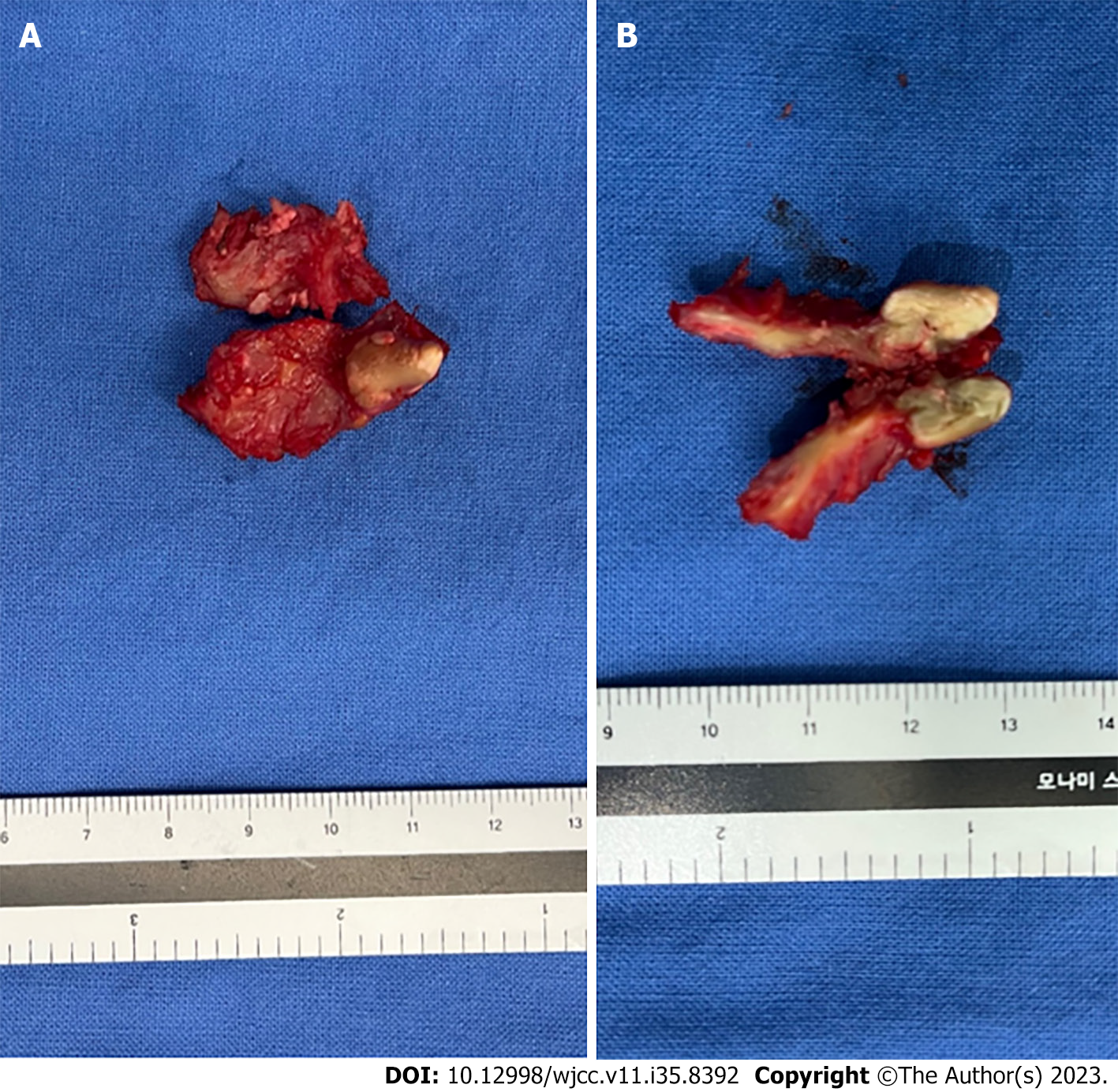
Figure 4 Intraoperative clinical photographs of the excised calcified cyst.
A firm, brown-colored, nodule-like mass originating from the ventral surface of the left side ligametum flavum was found. A: Ventral surface of the excised ligamentum flavum; B: Cross-section of the excised calcified cyst.
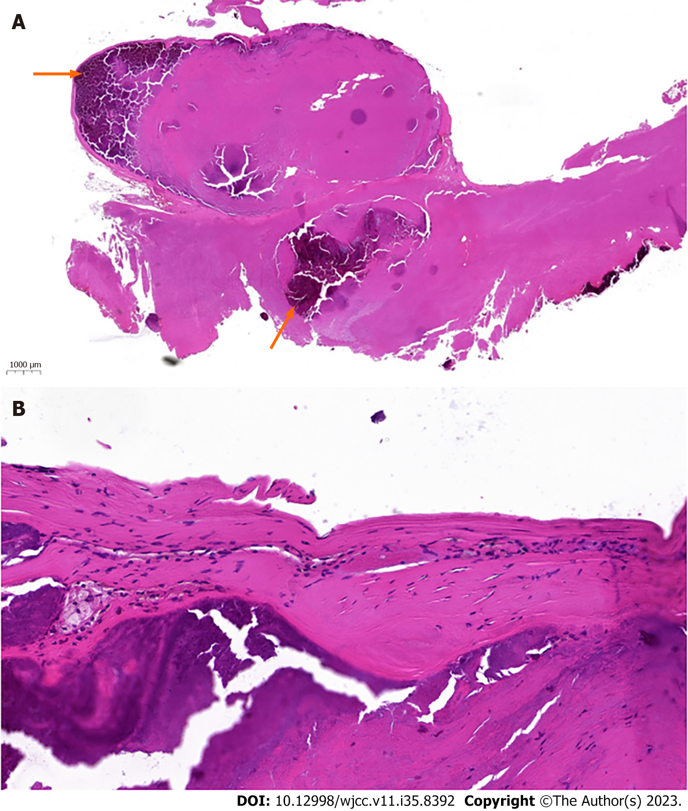
Figure 5 Microscopic images of the excised calcified cyst.
A: Low magnification (× 8) image showing a cyst in the ventral surface of ligamentum flavum with dark purple-colored calcified material in the cyst (arrow); B: Higher magnification (× 20) image showing no identifiable epithelial cell lining.
- Citation: Jung HY, Kim GU, Joh YW, Lee JS. Ankle and toe weakness caused by calcified ligamentum flavum cyst: A case report. World J Clin Cases 2023; 11(35): 8392-8398
- URL: https://www.wjgnet.com/2307-8960/full/v11/i35/8392.htm
- DOI: https://dx.doi.org/10.12998/wjcc.v11.i35.8392


