©The Author(s) 2023.
World J Clin Cases. Dec 16, 2023; 11(35): 8385-8391
Published online Dec 16, 2023. doi: 10.12998/wjcc.v11.i35.8385
Published online Dec 16, 2023. doi: 10.12998/wjcc.v11.i35.8385
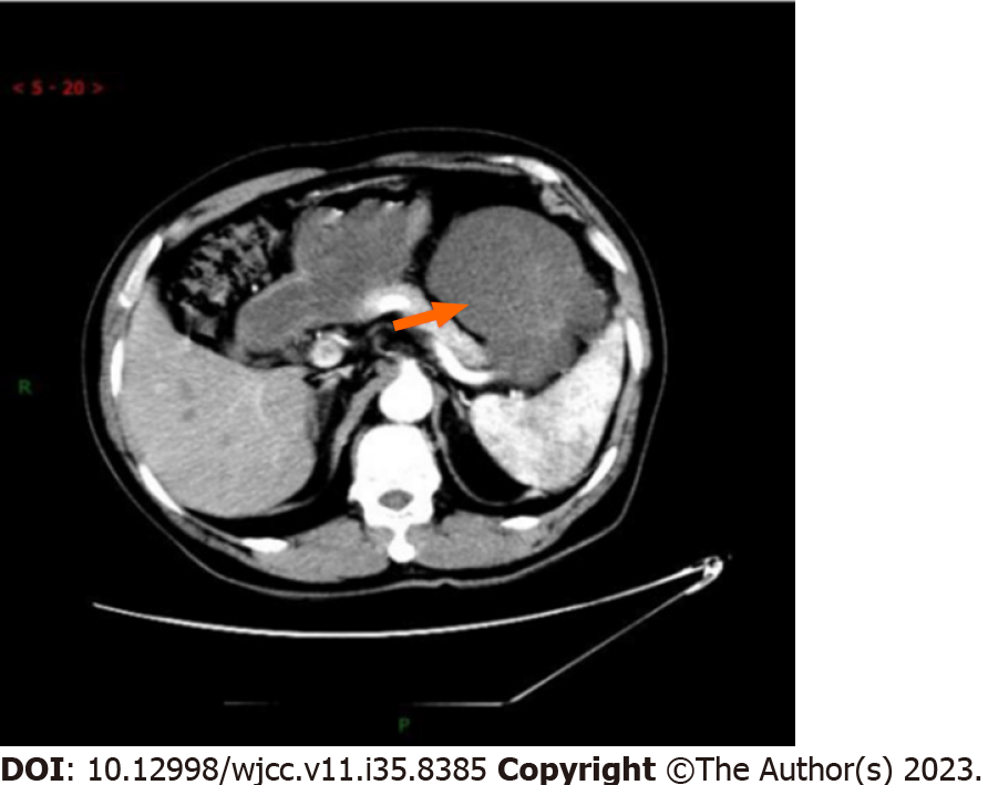
Figure 1 A 7.
2 cm × 9.7 cm cystic solid density shadow was seen in the left upper abdominal cavity.
Figure 2 Microscopic examination showed more spindle-shaped and short spindle-shaped cell proliferation, which arranged in interwoven and fascicles with moderate cell density.
A: HE, × 40; B and C: HE, × 200.
Figure 3 Computed tomography.
A: Multiple metastatic lesions in the abdominal cavity; B: Peritoneum; C: Abdominal wall.
Figure 4 Computed tomography.
A: After treatment with anlotinib combined with pablizumab, the abdominal cavity lesions; B: Were slightly larger and the peritoneal; C: Abdominal wall lesions were smaller than before.
- Citation: Wu RT, Zhang JC, Fang CN, Qi XY, Qiao JF, Li P, Su L. Anlotinib in combination with pembrolizumab for low-grade myofibroblastic sarcoma of the pancreas: A case report. World J Clin Cases 2023; 11(35): 8385-8391
- URL: https://www.wjgnet.com/2307-8960/full/v11/i35/8385.htm
- DOI: https://dx.doi.org/10.12998/wjcc.v11.i35.8385


