Copyright
©The Author(s) 2023.
World J Clin Cases. Dec 16, 2023; 11(35): 8379-8384
Published online Dec 16, 2023. doi: 10.12998/wjcc.v11.i35.8379
Published online Dec 16, 2023. doi: 10.12998/wjcc.v11.i35.8379
Figure 1 Thyroid ultrasound.
A: Left sided thyroid nodule oval shaped; B: With increased vascularity.
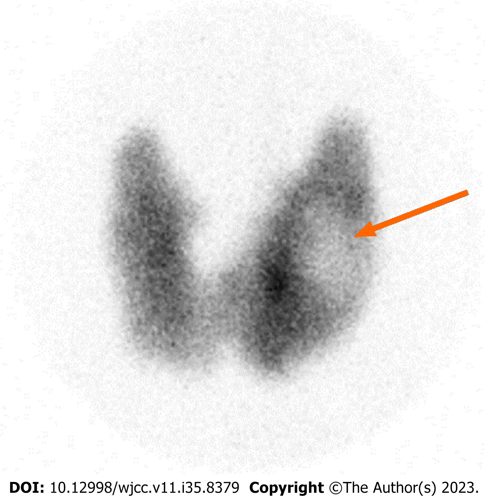
Figure 2 Technetium scan (Tc-99m) showing hyperthyroid gland with cold nodule (the arrow points to a cold nodule in the left thyroid lobe).
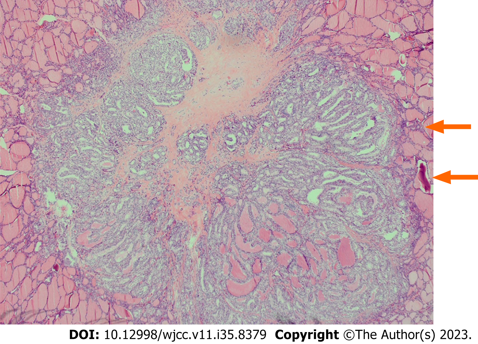
Figure 3 Papillary thyroid cancer showing neoplastic papillae (upper arrow) and psammoma bodies (lower arrow).
- Citation: Alzaman N. Multifocal papillary thyroid cancer in Graves’ disease: A case report. World J Clin Cases 2023; 11(35): 8379-8384
- URL: https://www.wjgnet.com/2307-8960/full/v11/i35/8379.htm
- DOI: https://dx.doi.org/10.12998/wjcc.v11.i35.8379


