©The Author(s) 2023.
World J Clin Cases. Dec 16, 2023; 11(35): 8364-8371
Published online Dec 16, 2023. doi: 10.12998/wjcc.v11.i35.8364
Published online Dec 16, 2023. doi: 10.12998/wjcc.v11.i35.8364
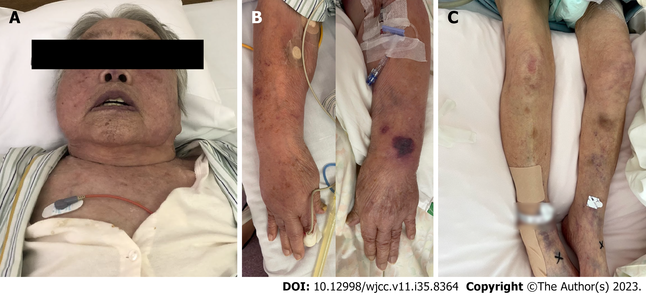
Figure 1 Physical findings on admission.
A: Facial oedema and cyanosis; B: Oedema in both upper limbs; C: No lower limb oedema.
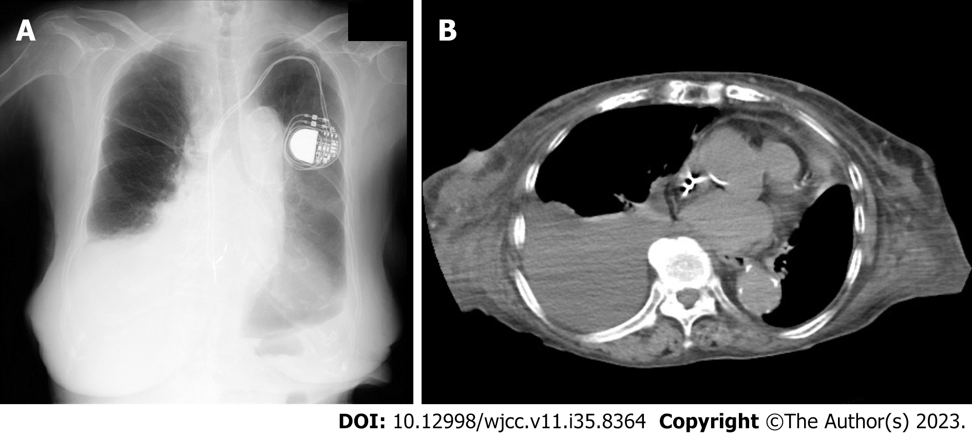
Figure 2 Imaging examinations on admission.
A: Chest radiograph; B: Chest computed tomography image. Both images show a massive right pleural effusion.
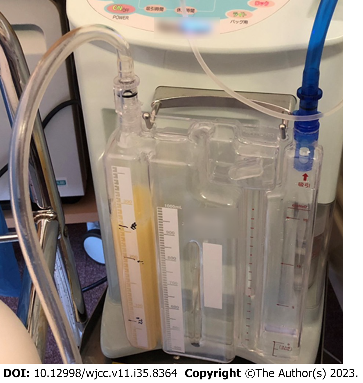
Figure 3 Chest tube drainage persistently shows a milky white pleural effusion.
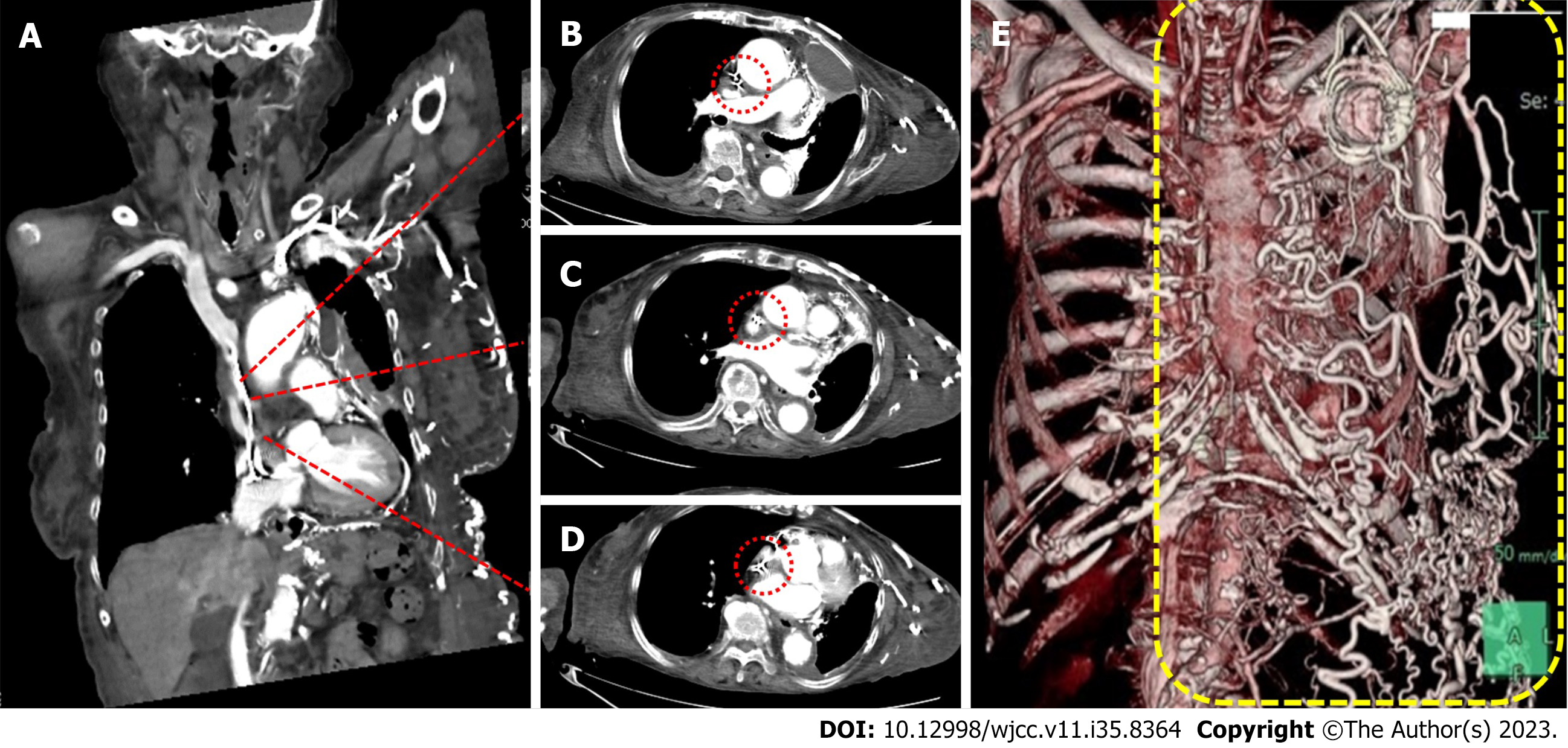
Figure 4 Contrast-enhanced computed tomography of the superior vena cava.
A: Coronal section of the upper body; B-D: Transverse sections of the upper body; the dashed red line represents the superior vena cava (the most stenotic site; D); E: 3D volume-rendered image of the collateral circulation (inside the yellow-dashed rectangle).
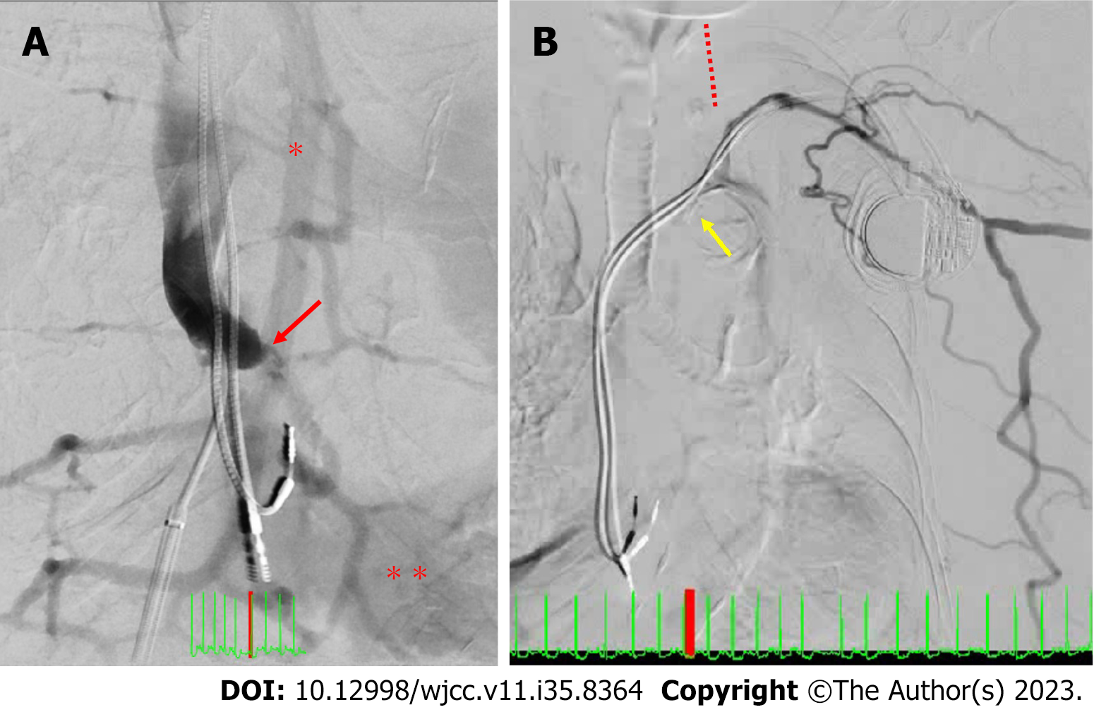
Figure 5 Digital subtraction venography before venoplasty.
A: Digital subtraction venography (DSV) for the superior vena cava (SVC); B: DSV from the left forearm. Red arrow indicates the SVC occlusion site; One asterisk represents the azygos vein; Two asterisks represent the right ventricle; Yellow arrow indicates an occluded innominate vein; Red dashed line represents a non-contrasted left internal jugular vein.
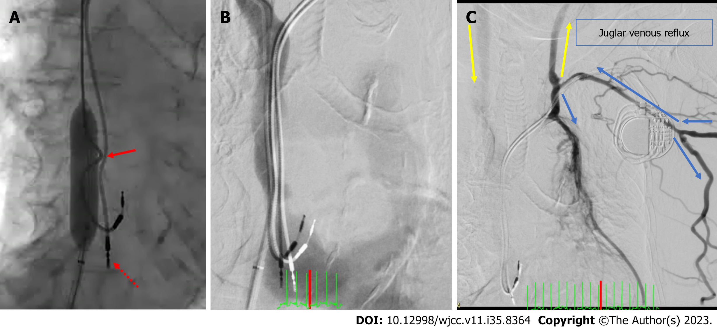
Figure 6 Digital subtraction venography before and after venoplasty.
A: Balloon venoplasty for the superior vena cava (SVC); B: Digital subtraction venography (DSV) from the SVC after venoplasty; C: DSV from the left forearm after venoplasty. The red dashed arrow indicates the dislodged right ventricular lead. The red arrow shows the bent right atrium (RA) lead caused by balloon expansion; the blood moved directly into the RA; The blue arrow shows the direction of the blood flow. Some blood flowed into the collateral circulation in the abdominal wall; The yellow arrow shows the jugular venous reflux.
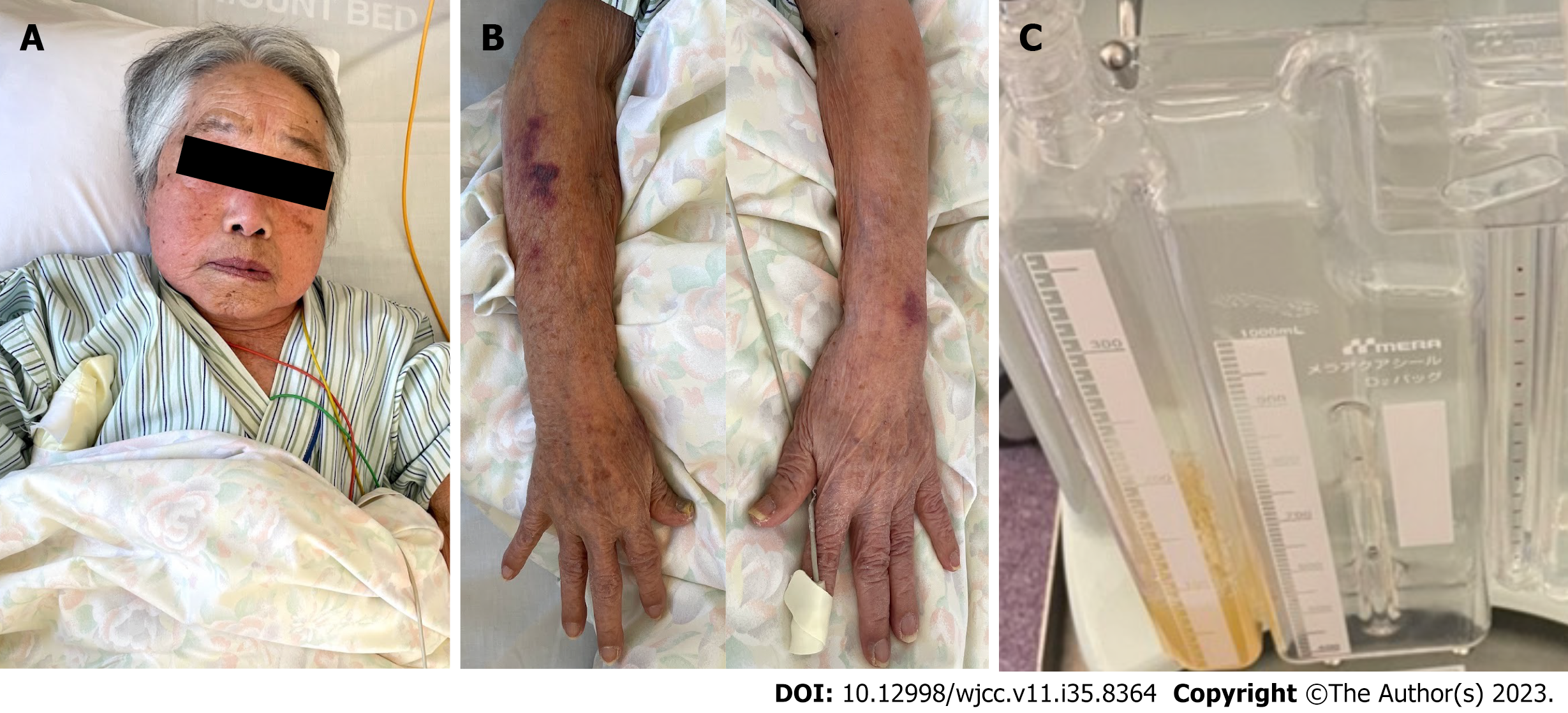
Figure 7 Physical findings after treatment.
A: Facial oedema and cyanosis improved after venoplasty; B: Oedema in both the upper limbs disappeared after venoplasty; C: Chylothorax improved, and the pleural effusion turned a transparent yellow.
- Citation: Yamamoto S, Kamezaki M, Ooka J, Mazaki T, Shimoda Y, Nishihara T, Adachi Y. Balloon venoplasty for disdialysis syndrome due to pacemaker-related superior vena cava syndrome with chylothorax post-bacteraemia: A case report. World J Clin Cases 2023; 11(35): 8364-8371
- URL: https://www.wjgnet.com/2307-8960/full/v11/i35/8364.htm
- DOI: https://dx.doi.org/10.12998/wjcc.v11.i35.8364


