Copyright
©The Author(s) 2023.
World J Clin Cases. Dec 6, 2023; 11(34): 8205-8211
Published online Dec 6, 2023. doi: 10.12998/wjcc.v11.i34.8205
Published online Dec 6, 2023. doi: 10.12998/wjcc.v11.i34.8205
Figure 1 A 58-year-old woman presented with Sister Mary Joseph's nodule.
A and B: A 2-cm hard, localized, and painless nodule with erosions observed in the umbilical region; C: Positron emission tomography (PET) demonstrates increased 18F-fluorodeoxyglucose uptake in the umbilicus, right supraclavicular fossa, and bilateral lung fields (red arrow); D: PET-computed tomography scan revealed increased 18F-fluorodeoxyglucose uptake in the umbilicus (yellow arrow), but there were no nodules or abdominal/pelvic fluid suggesting tumor metastasis to the peritoneum.
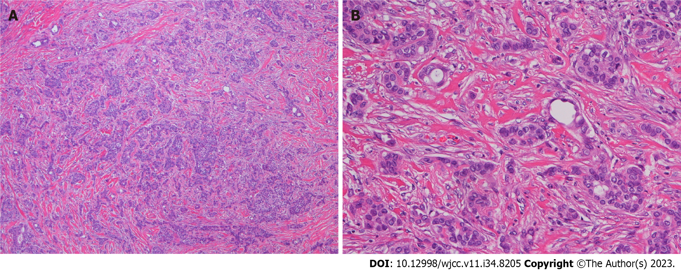
Figure 2 Pathology of the metastatic specimens.
A: Morphology (Hematoxylin and eosin, × 40); B: Morphology (Hematoxylin and eosin, × 200) confirming an adenocarcinoma in a foamy, tubular arrangement in metastatic lesions.
Figure 3 Perioperative images of tumor resection and immediate abdominal wall reconstruction.
A: Tumor excision with a 15 mm horizontal margin, encompassing a combined resection of the peritoneum and the falciform ligament; B: Image showing no exposure of the tumor to the abdominal cavity; C: Macroscopic image of the resected tissue; D–F: Abdominal wall reconstruction using a component-separation technique.
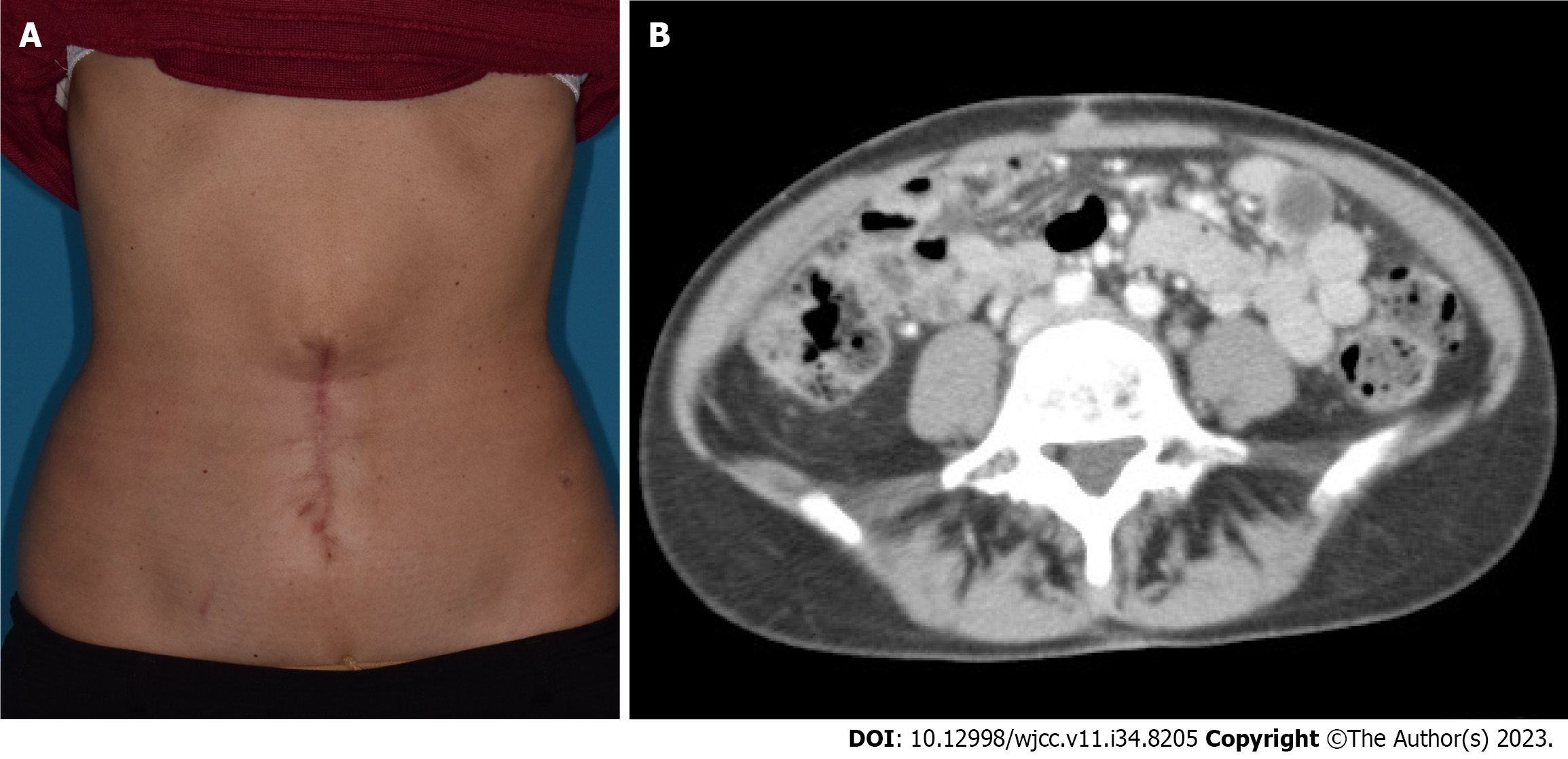
Figure 4 Five-year postoperative images.
A: No evident local recurrence in the umbilical region; B: Computed tomography scan shows no nodules, suggesting tumor recurrence in the umbilical region or metastasis to the peritoneum in the abdominal cavity.
- Citation: Kanayama K, Tanioka M, Hattori Y, Iida T, Okazaki M. Long-term survival of the Sister Mary Joseph nodule originating from breast cancer: A case report. World J Clin Cases 2023; 11(34): 8205-8211
- URL: https://www.wjgnet.com/2307-8960/full/v11/i34/8205.htm
- DOI: https://dx.doi.org/10.12998/wjcc.v11.i34.8205


