©The Author(s) 2023.
World J Clin Cases. Nov 26, 2023; 11(33): 8084-8088
Published online Nov 26, 2023. doi: 10.12998/wjcc.v11.i33.8084
Published online Nov 26, 2023. doi: 10.12998/wjcc.v11.i33.8084
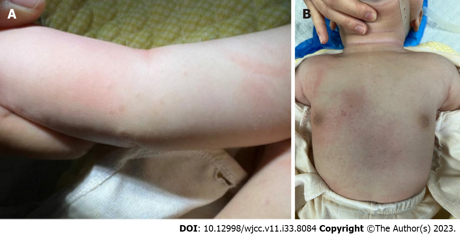
Figure 1 Clinical manifestation.
A: Papules of different sizes scattered on the upper limbs that are reddish and light brown on the surface and firm in texture; B: Scattered pale white pitting macules on the chest and abdomen, resembling scar-like changes in appearance, with normal texture.
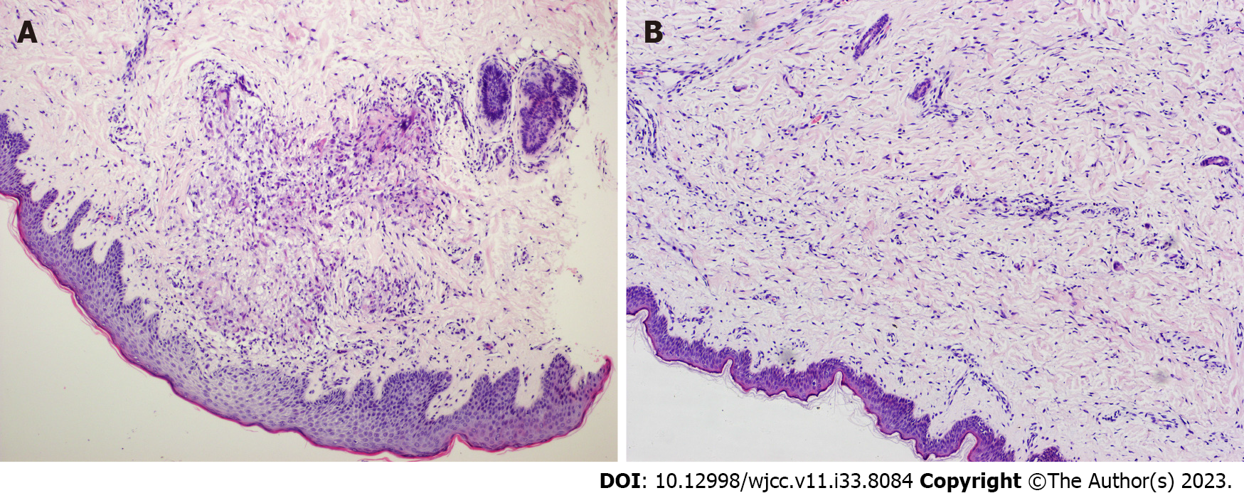
Figure 2 Images in pathology (hematoxylin and eosin stain).
A: Pathological findings of papular lesions (epithelioid nodular granuloma annular (GA); hematoxylin and eosin (HE) ×100): Focal mild thickening of the epidermis and nested infiltration of lymphocytes, histiocytes, and multinucleated giant cells in the superficial and middle dermis, with some collagen disarrangement; B: Pathological findings of scar-like lesions in the depressions (scattered histiocytic infiltrative GA; HE ×100): The epidermis is approximately normal, and lymphocytes and histiocytes have infiltrated between the collagen fibers and around blood vessels in the superficial middle dermis. Some collagen fibers have degenerated, and the intervals are widened.
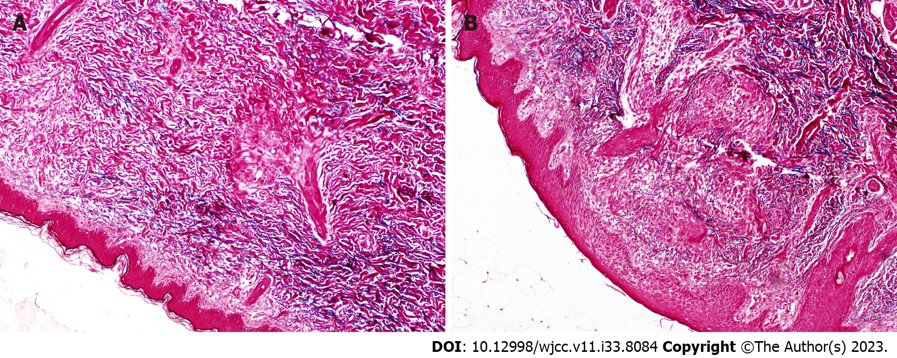
Figure 3 Images in pathology (elastic fiber stain).
A: Elastic fiber staining (×100) in scar-like lesions in the depressions: Elastic fibers have decreased or disappeared; B: Staining of elastic fibers in papular lesions (×100): Elastic fibers have decreased or are absent.
- Citation: Zhang DY, Zhang L, Yang QY, Li J, Jiang HC, Xie YC, Shu H. Generalized granuloma annulare in an infant clinically manifested as papules and atrophic macules: A case report. World J Clin Cases 2023; 11(33): 8084-8088
- URL: https://www.wjgnet.com/2307-8960/full/v11/i33/8084.htm
- DOI: https://dx.doi.org/10.12998/wjcc.v11.i33.8084


