©The Author(s) 2023.
World J Clin Cases. Nov 16, 2023; 11(32): 7865-7871
Published online Nov 16, 2023. doi: 10.12998/wjcc.v11.i32.7865
Published online Nov 16, 2023. doi: 10.12998/wjcc.v11.i32.7865
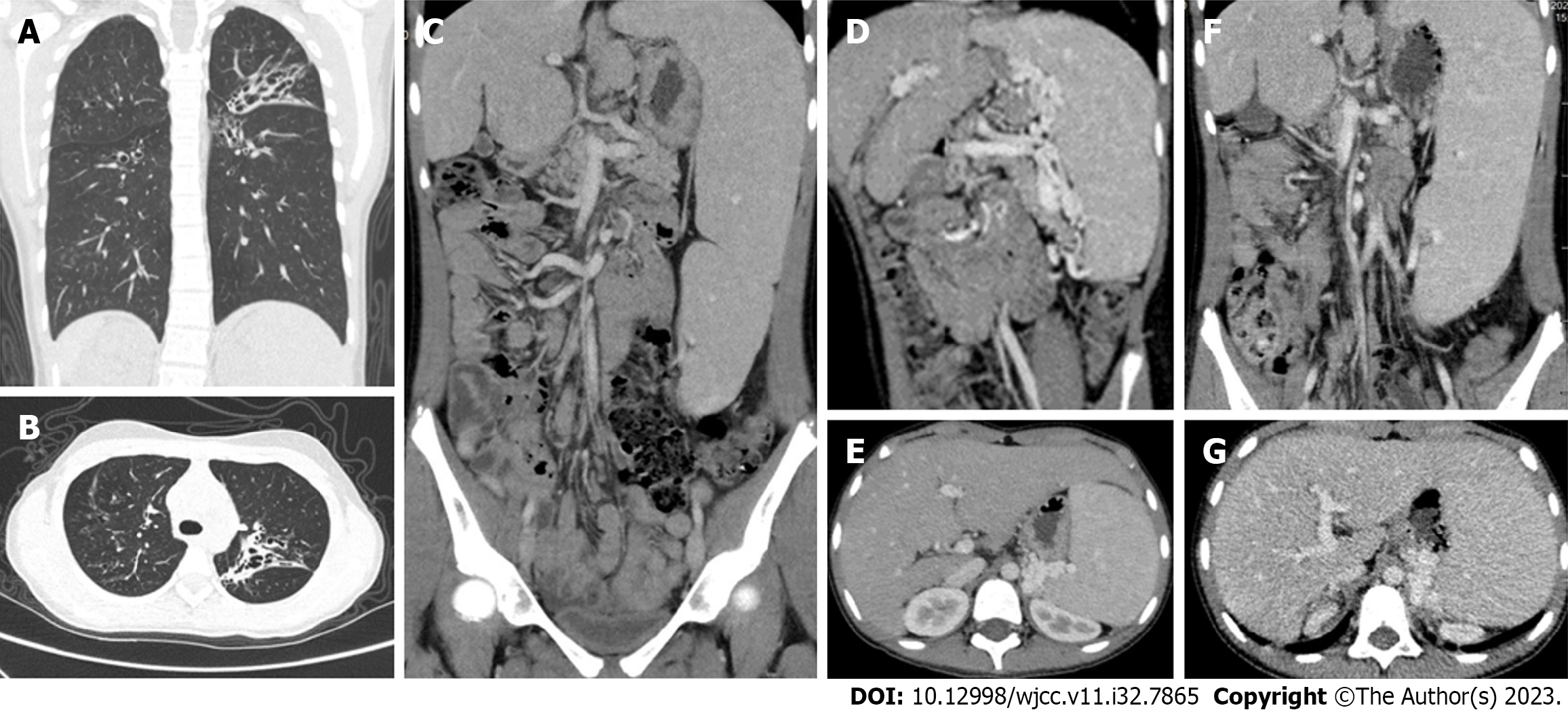
Figure 1 Chest and abdominal computed tomography.
A and B: Chest computed tomography (CT) showed bronchial dilatation and flocculent shadow with multiple cystic translucency in both lungs; C-E: Preoperative whole abdominal enhanced CT showed splenomegaly, multiple tortuous dilated vessels at the splenic hilum, irregular liver morphology and pancreatic atrophy; F and G: 17-month postoperative whole abdomen enhanced CT showed irregular liver morphology, splenomegaly, multiple tortuous dilated vessels at the splenic hilum, and pancreatic atrophy.
Figure 2 CFTR gene sequencing.
A: The mother of the proband was a carrier of CFTR gene c.233_c.234insT heterozygous mutation compared with the standard sequence; B: The father of the proband carried CFTR gene c.2909G>A/p.Gly970Asp heterozygous mutation.
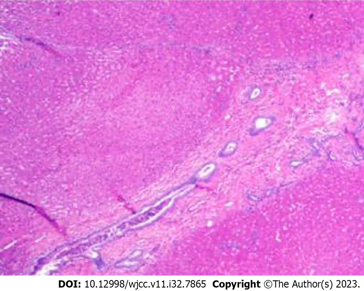
Figure 3 Pathology of liver biopsy.
Pathological findings of the liver biopsy showed poorly developed, dilated or absent portal vein branches.
- Citation: Zhang LJ, Liu XY, Chen TF, Xu ZY, Yin HJ. Type II Abernethy malformation with cystic fibrosis in a 12-year-old girl: A case report. World J Clin Cases 2023; 11(32): 7865-7871
- URL: https://www.wjgnet.com/2307-8960/full/v11/i32/7865.htm
- DOI: https://dx.doi.org/10.12998/wjcc.v11.i32.7865


