©The Author(s) 2023.
World J Clin Cases. Nov 6, 2023; 11(31): 7663-7672
Published online Nov 6, 2023. doi: 10.12998/wjcc.v11.i31.7663
Published online Nov 6, 2023. doi: 10.12998/wjcc.v11.i31.7663
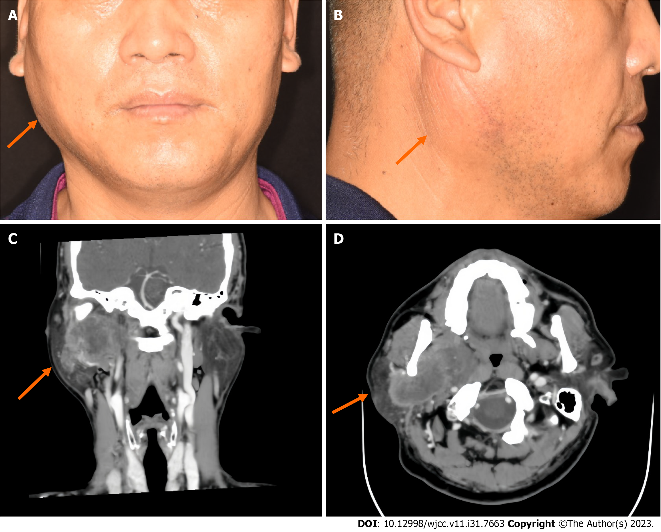
Figure 1 Preoperative photographs and enhanced computed tomography images of the patient.
A: Front; B: Side; C: Computed tomography of the right parotid gland revealed a mass in its deep lobe that also extended into the parapharynx; D: There was obvious uneven enhancement, with local ring enhancement.
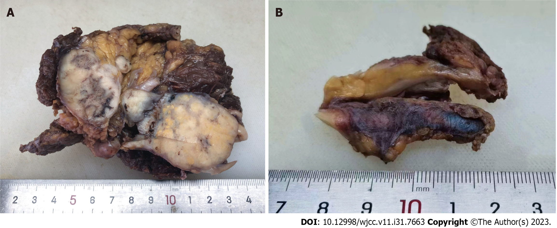
Figure 2 Gross findings of the excised specimen.
A: The cut surface of the mass was gray-white, solid, and tough; B: Thickened venous tissue was observed.
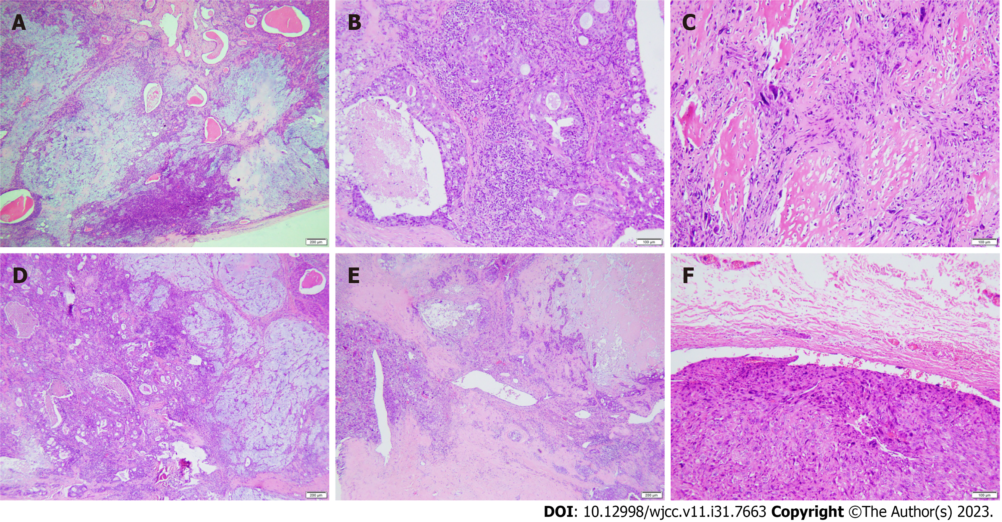
Figure 3 Histological features of carcinosarcoma (hematoxylin and eosin staining).
A: The pleomorphic adenoma region had a rich tissue structure and mild cell morphological changes; B: In the salivary duct carcinoma area, comedo-like necrosis was seen in the center of the solid epithelial mass; C: In the sarcomatous area, the cells showed obvious atypia and abundant bone matrix formation around the cells; D: Pleomorphic adenoma (right) mixed with salivary duct carcinoma (left); E: Salivary duct carcinoma (right) mixed with osteosarcoma (left); F: A tumor thrombus was found in the venous lumen.
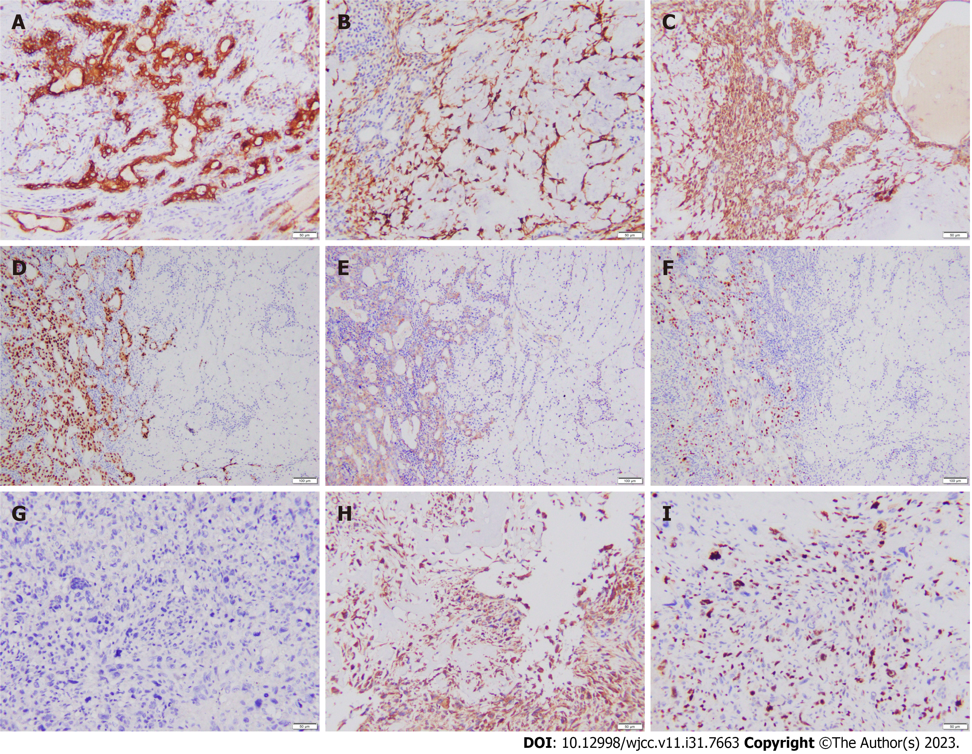
Figure 4 Immunohistochemical results.
A-C: The tumor epithelium in the pleomorphic adenoma area was positive for CK7 (A), calponin (B), and CK5/6 (C); D-F: In the mixed area of pleomorphic adenoma (right) and salivary duct carcinoma (left), the tumor epithelium in the salivary duct carcinoma area was positive for AR (D) and HER2 (E), and the Ki-67 proliferation index in the salivary duct carcinoma area was higher than that in the pleomorphic adenoma area (F); G: PCK was negative in osteosarcoma tumor cells; H: Cells in the osteosarcoma area were positive for vimentin; I: The Ki-67 proliferation index of tumor cells in the osteosarcoma area was approximately 50%.
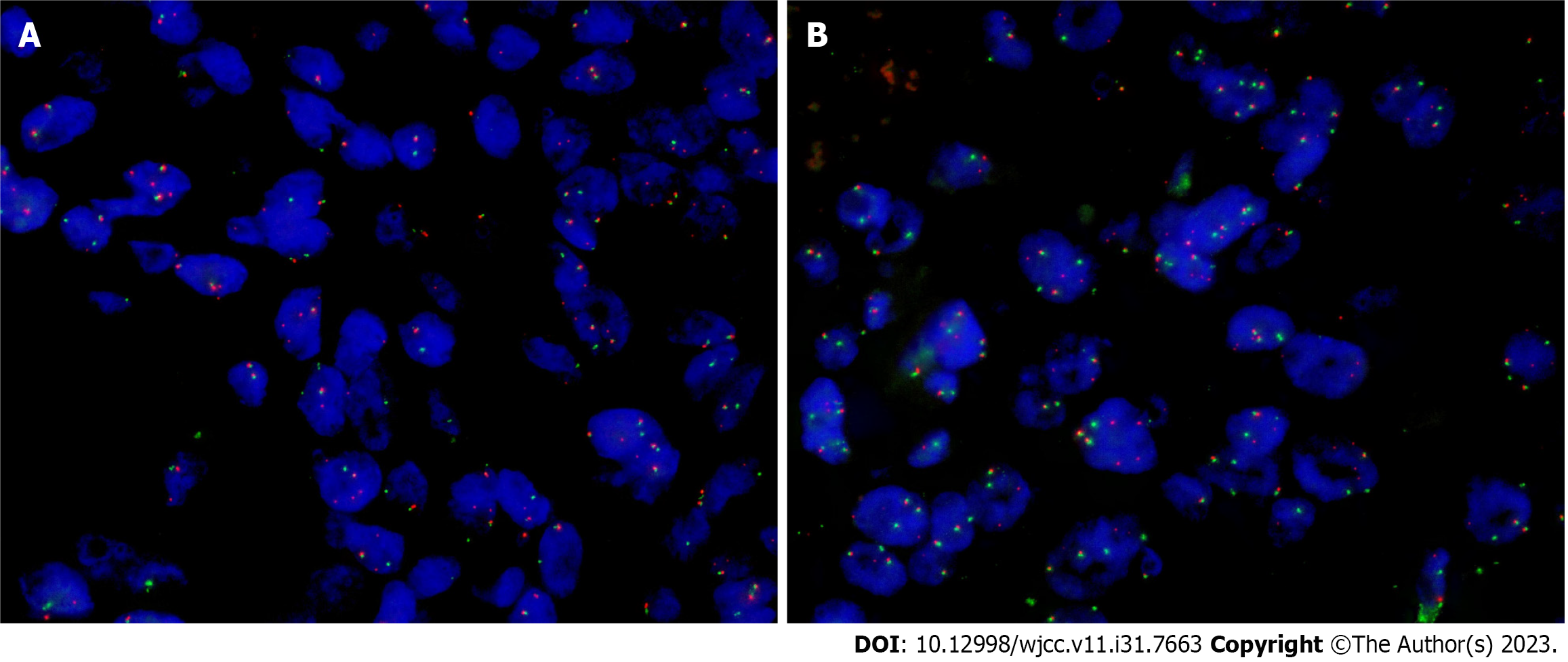
Figure 5 Fluorescence in situ hybridization detection of PLAG1 gene rearrangement in tumor tissue.
A: PLAG1 rearrangement was observed in pleomorphic adenomas by fluorescence in situ hybridization (FISH); B: FISH assay of the salivary duct carcinoma component showed rearrangement of PLAG1.
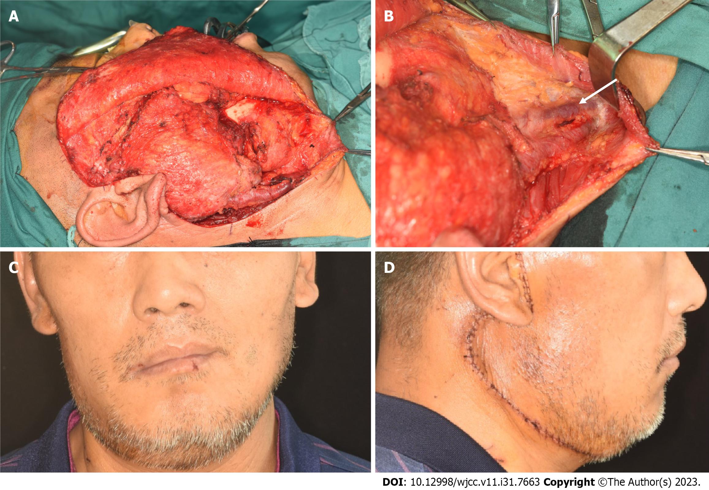
Figure 6 Intraoperative and one-week-postoperative photographs.
A: Location of the mass; B: Thickened and hardened external jugular veins; C: Front; D: Side.
- Citation: Tang YY, Zhu GQ, Zheng ZJ, Yao LH, Wan ZX, Liang XH, Tang YL. Carcinosarcoma of the deep lobe of the parotid gland in the parapharyngeal region: A case report. World J Clin Cases 2023; 11(31): 7663-7672
- URL: https://www.wjgnet.com/2307-8960/full/v11/i31/7663.htm
- DOI: https://dx.doi.org/10.12998/wjcc.v11.i31.7663


