Copyright
©The Author(s) 2023.
World J Clin Cases. Oct 16, 2023; 11(29): 7127-7135
Published online Oct 16, 2023. doi: 10.12998/wjcc.v11.i29.7127
Published online Oct 16, 2023. doi: 10.12998/wjcc.v11.i29.7127
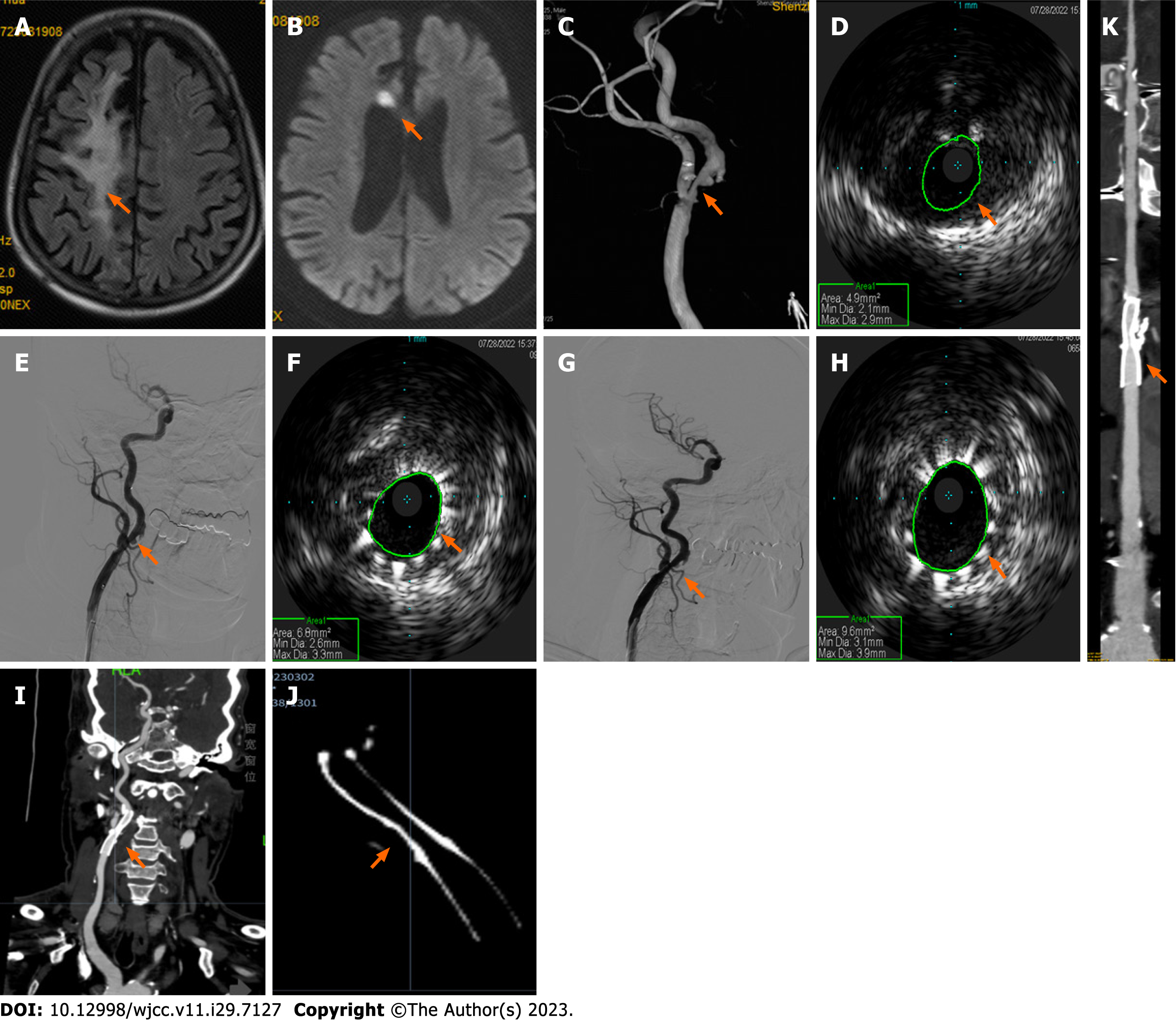
Figure 1 Neuroimaging results of patient one during intravascular ultrasound-assisted carotid artery stenting.
A and B: Brain magnetic resonance imaging demonstrates a paraventricular white-matter lesion in the right frontal lobe (A, arrow) on fluid-attenuated inversion recovery imaging, and a small acute infarction at the genu of the corpus callosum on diffusion weighted imaging (B, arrow); C: Angiography shows 70% stenosis of the initial segment of the right internal carotid artery (ICA) (arrow); D and E: Preoperative intravascular ultrasound (IVUS) displays severe stenosis with plaque formation and obvious calcification under plaque (D, arrow) and a well-positioned stent (E, arrow); F-H: Subsequent IVUS imaging confirmed improvement of the narrowest part (F, arrow), thus post-stent balloon dilatation was performed. Postoperative angiography shows a right ICA residual stenosis of 20% (G, arrow), and IVUS confirmed good stent expansion and adherence (H, arrow); I-K: Computed tomography angiography six months postoperatively revealed mild in-stent stenosis in the right initial segment of the ICA (I, multiplanar reconstruction; J, amplification imaging with adjusting parameters; K, curve planar reformation; arrows).
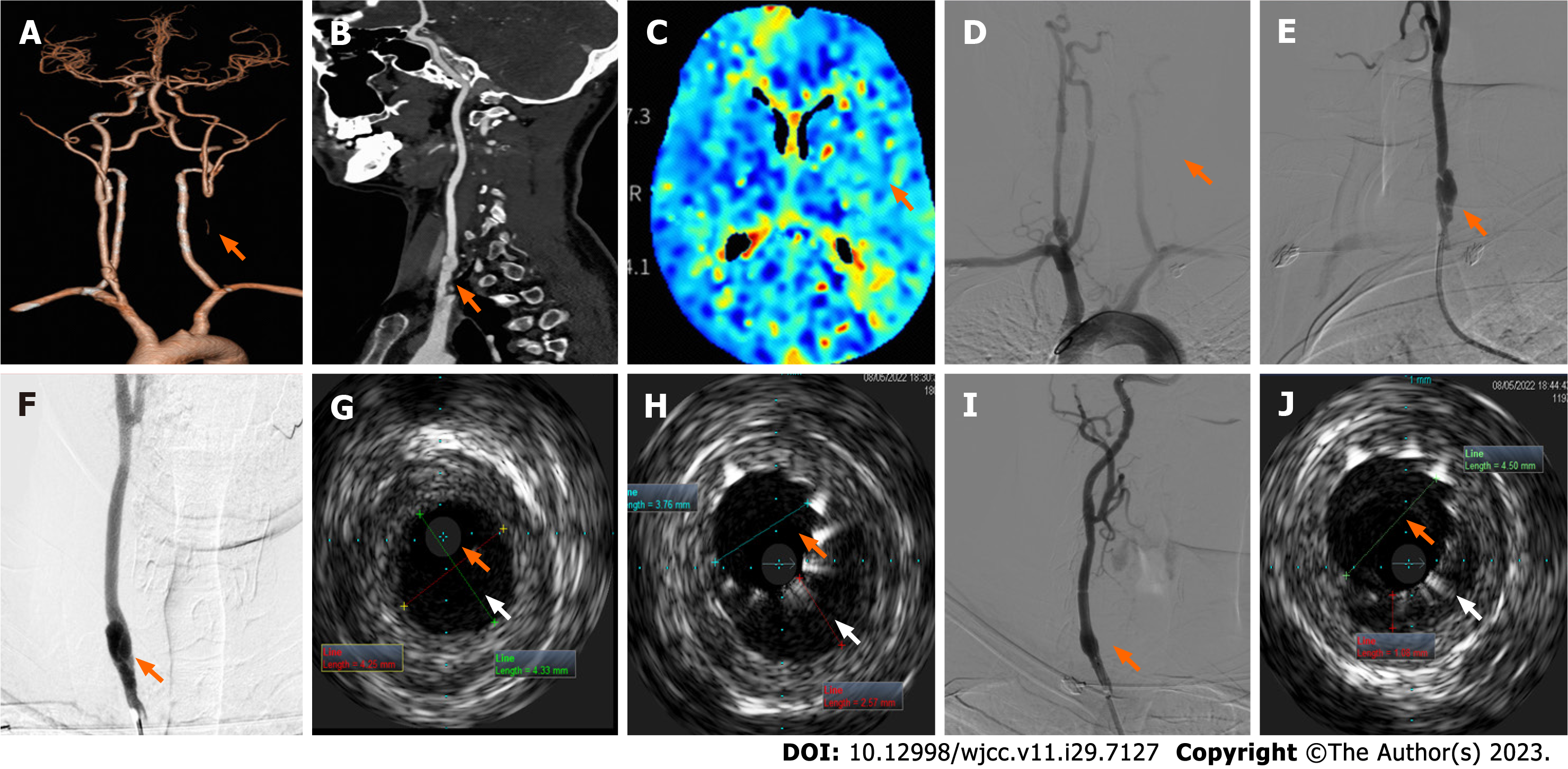
Figure 2 Neuroimaging results of patient two during intravascular ultrasound-assisted carotid artery stenting.
A and B: Head and neck computed tomography angiography showing left common carotid artery (CCA) occlusion (A, arrow) and local bulging in the inferior segment of the right CCA with strip-shaped, low-density shadow (B, arrow); C: Brain computed tomography perfusion images showed that blood flow perfusion in the left frontal lobe, parietal lobe, right temporal lobe, and insula was lower than that on the opposite side (arrow); D-F: Digital subtraction angiography shows the initial segment of the left CCA occlusion (D, arrow) and the initial segment of the right CCA dissection (posteroanterior film, E; left oblique film, F); G: Preoperative intravascular ultrasonography (IVUS) probe check dissection; H: IVUS images after stent placement show insufficient true lumen (orange arrow) with visible intimal flaps, and a relatively large pseudo-lumen (white arrow) with a large intermural hematoma; I: Angiography after post-balloon dilatation showed a smooth intra-arterial lumen; J: Postoperative IVUS images showed that the true lumen enlarged (orange arrow), the pseudo-lumen diminished (white arrow), and confirmed satisfactory stent expansion and adherence.
- Citation: Fu PC, Wang JY, Su Y, Liao YQ, Li SL, Xu GL, Huang YJ, Hu MH, Cao LM. Intravascular ultrasonography assisted carotid artery stenting for treatment of carotid stenosis: Two case reports. World J Clin Cases 2023; 11(29): 7127-7135
- URL: https://www.wjgnet.com/2307-8960/full/v11/i29/7127.htm
- DOI: https://dx.doi.org/10.12998/wjcc.v11.i29.7127


