©The Author(s) 2023.
World J Clin Cases. Jul 26, 2023; 11(21): 5160-5166
Published online Jul 26, 2023. doi: 10.12998/wjcc.v11.i21.5160
Published online Jul 26, 2023. doi: 10.12998/wjcc.v11.i21.5160
Figure 1 Ultrasound image of the uterus.
A: The circular area indicates the endometrial area; B: Endometrial tumor thickness 3.02 cm; C: Uterine tumor 5.2 cm.
Figure 2 Computed tomography of endometrial cancer.
A: Sagittal view, B: Coronal view; C: Axial view. Hypointensity represented the tumor site.
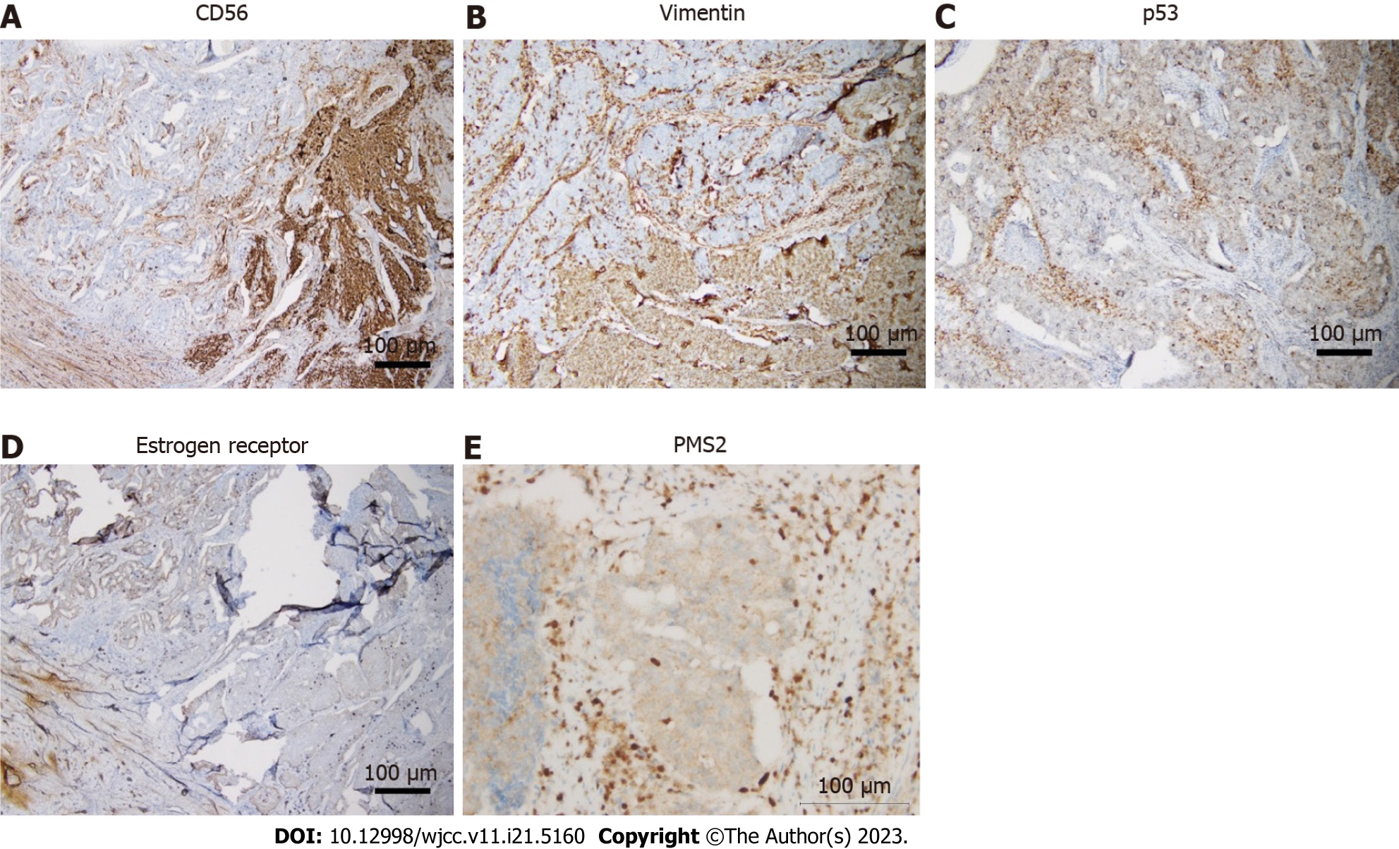
Figure 3 Immunohistochemistry of endometrial neuroendocrine carcinoma.
A: CD56; B: Vimentin; C: p53; D: Estrogen receptor; E: PMS2. Scale bar = 100 μm.
- Citation: Siu WYS, Hong MK, Ding DC. Neuroendocrine carcinoma of the endometrium concomitant with Lynch syndrome: A case report. World J Clin Cases 2023; 11(21): 5160-5166
- URL: https://www.wjgnet.com/2307-8960/full/v11/i21/5160.htm
- DOI: https://dx.doi.org/10.12998/wjcc.v11.i21.5160


