©The Author(s) 2023.
World J Clin Cases. Jul 6, 2023; 11(19): 4723-4728
Published online Jul 6, 2023. doi: 10.12998/wjcc.v11.i19.4723
Published online Jul 6, 2023. doi: 10.12998/wjcc.v11.i19.4723
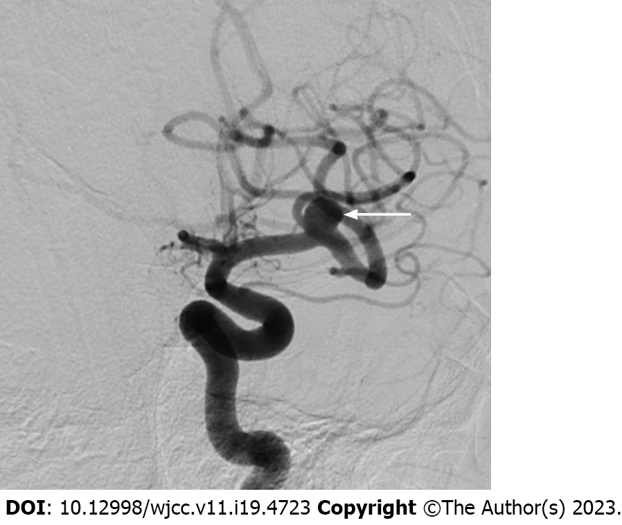
Figure 1 Left internal carotid artery angiography.
Preoperative image shows a small aneurysm (arrow) at the left middle cerebral artery bifurcation with an unfavorable dome-to-neck ratio.
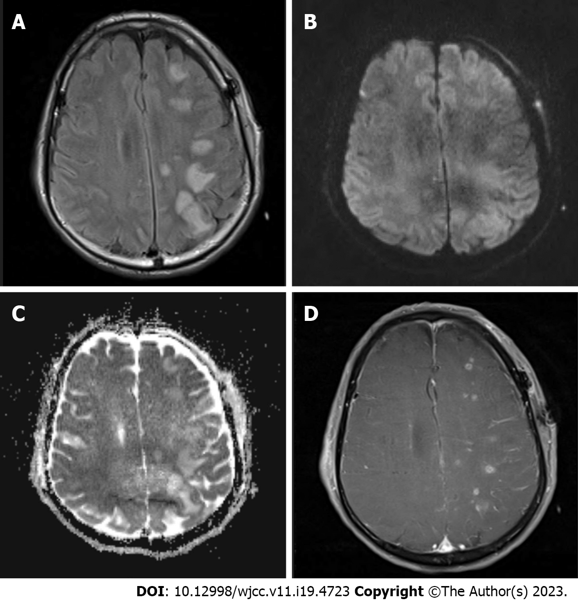
Figure 2 Magnetic resonance imaging obtained immediately following deterioration of consciousness A: Axial fluid attenuation inversion recovery image shows extensive hyperintense lesions predominantly in the cortex and subcortical white matter of the left frontoparietal lobe; B: Diffusion-weighted image shows that lesions are iso to hyperintense without any signs of restricted diffusion; C: Apparent diffusion coefficient maps show that lesions are hyperintense, indicating vasogenic edema; D: On the contrast-enhanced three-dimensional T1-weighted image, lesions show patchy enhancement.
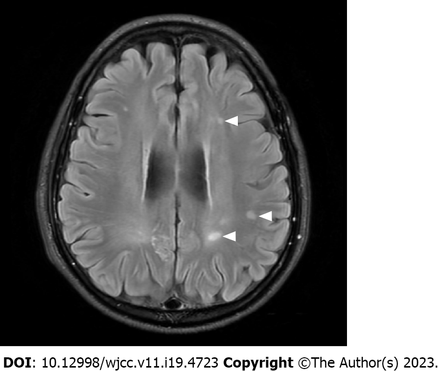
Figure 3 Follow-up magnetic resonance imaging.
Fluid attenuation inversion recovery image performed at one year after clipping surgery shows that several lesions (arrow heads) in the subcortical white matter persisted, despite being reduced in size.
- Citation: Hwang J, Cho WH, Cha SH, Ko JK. Posterior reversible encephalopathy syndrome following uneventful clipping of an unruptured intracranial aneurysm: A case report. World J Clin Cases 2023; 11(19): 4723-4728
- URL: https://www.wjgnet.com/2307-8960/full/v11/i19/4723.htm
- DOI: https://dx.doi.org/10.12998/wjcc.v11.i19.4723


