Copyright
©The Author(s) 2023.
World J Clin Cases. Jun 26, 2023; 11(18): 4438-4445
Published online Jun 26, 2023. doi: 10.12998/wjcc.v11.i18.4438
Published online Jun 26, 2023. doi: 10.12998/wjcc.v11.i18.4438
Figure 1 Preoperative findings.
Image showing a normal skin-colored mass measuring 2.5 cm × 3.5 cm. The patient did not show symptoms of tenderness, swelling, infection, or any abnormal sensation of the skin surface. A: Anterior view; B: Lateral view.
Figure 2 Preoperative facial animation test results.
No abnormalities of facial expression are observed.
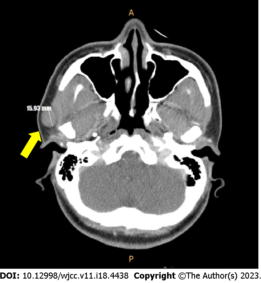
Figure 3 Facial computed tomography scan (axial view) findings.
Image showing a well-encapsulated low-density mass (yellow arrow) adherent to the inferior portion of the superficial lobe of the parotid gland.
Figure 4 Photograph showing superficial parotidectomy using a modified-Blair incision.
The incision commences from the anterior and superior aspects of the tragus along the preauricular skin crease and extends to the lower earlobe and postauricular area (blue arrow). The drawn incision marking line extends up to the mandibular border of the chin but not incised in operation (red arrow).
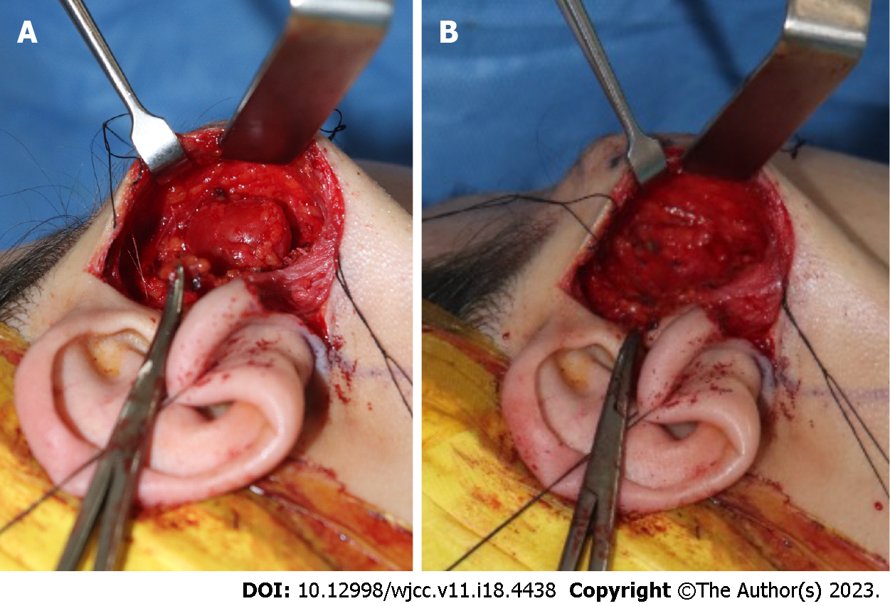
Figure 5 Photographs showing intraoperative findings.
A: Intracapsular enucleation of the schwannoma with preservation of the facial nerve fibers; B: Post-excision photograph showing that branches of the facial nerve coursing through the parotid gland are intact.
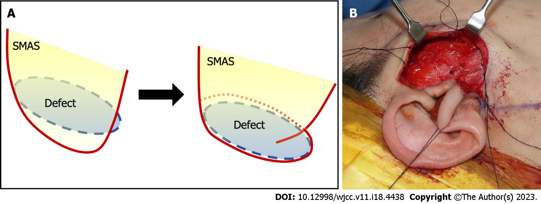
Figure 6 Photographs showing the superficial musculoaponeurotic system folding procedure.
A: Image showing folding of the lateral portion of the superficial musculoaponeurotic system (SMAS) layer (approximately 2 cm) to fill the post-parotidectomy defect; B: The defect is completely covered after being filled with the folded SMAS (blue circle). SMAS: Superficial musculoaponeurotic system.
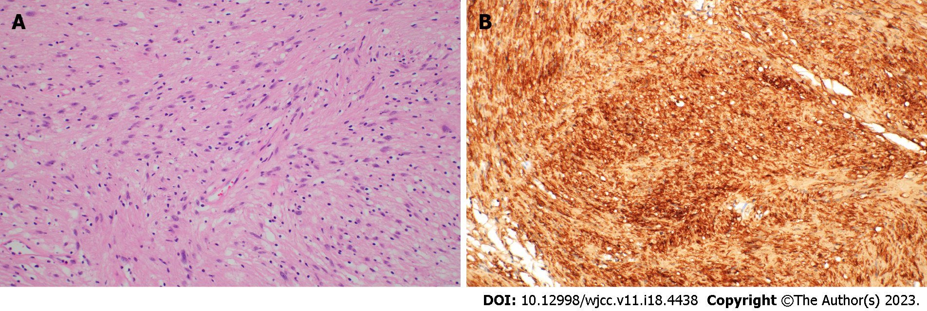
Figure 7 Histopathological findings of the resected specimen.
A: The tumor is composed of spindle cells with bland-looking nuclei and abundant cytoplasm (hematoxylin & eosin, × 200); B: Tumor showing diffuse strong immunoreactivity for the S100 protein (× 200).
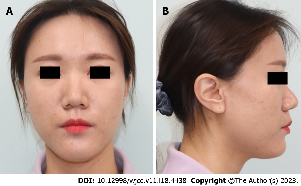
Figure 8 Photographs obtained at the 2-mo postoperative follow-up.
The incision scar is unnoticeable, and no specific complication is detected. The right cheek contour is maintained, and no differences are observed compared with the contralateral side. A: Anterior view; B: Lateral view.
- Citation: Nam HJ, Choi HJ, Byeon JY, Wee SY. Removal of unexpected schwannoma with superficial parotidectomy using modified-Blair incision and superficial musculoaponeurotic system folding: A case report. World J Clin Cases 2023; 11(18): 4438-4445
- URL: https://www.wjgnet.com/2307-8960/full/v11/i18/4438.htm
- DOI: https://dx.doi.org/10.12998/wjcc.v11.i18.4438


