Copyright
©The Author(s) 2023.
World J Clin Cases. Jun 16, 2023; 11(17): 4117-4122
Published online Jun 16, 2023. doi: 10.12998/wjcc.v11.i17.4117
Published online Jun 16, 2023. doi: 10.12998/wjcc.v11.i17.4117
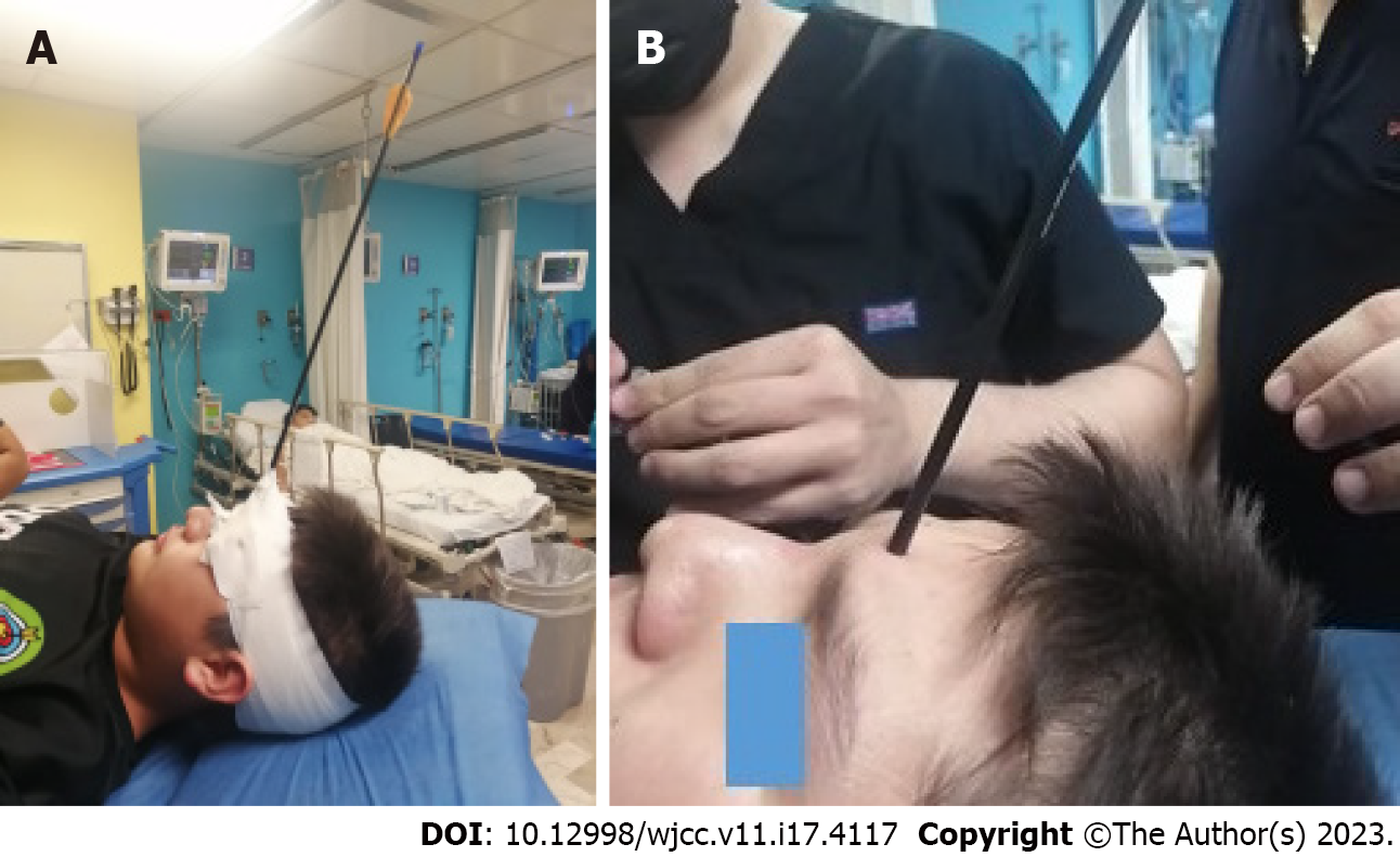
Figure 1 Photographs of the patient with a foreign body penetrating the frontal region.
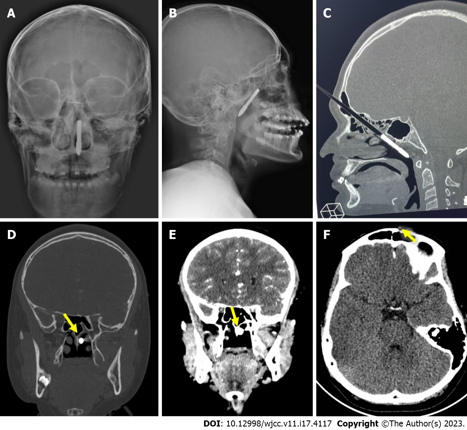
Figure 2 Radiological findings of the patient with a foreign body in the frontal region.
A and B: Frontal and lateral skull X-rays show a foreign body (arrow) with an entry site in the left frontal region penetrating the posterior pharynx through the ethmoid cells; C: A simple computerized tomography (CT) scan of the skull shows the inferior oblique trajectory through the ethmoid cells of the foreign body (arrow) with the distal end in the nasopharynx; D and E: Cranial coronal CT-scan and angio-CT showing the foreign body (yellow arrows) medial to the middle and inferior turbinates with no evidence of nasal septum fracture or lesions in vascular structures; F: Postoperative simple cranial CT scan shows a fracture of the external wall of the left frontal bone (yellow arrow).
- Citation: Rodríguez-Ramos A, Zapata-Castilleja CA, Treviño-González JL, Palacios-Saucedo GC, Sánchez-Cortés RG, Hinojosa-Amaya LG, Nieto-Sanjuanero A, de la O-Cavazos M. Frontal penetrating arrow injury: A case report. World J Clin Cases 2023; 11(17): 4117-4122
- URL: https://www.wjgnet.com/2307-8960/full/v11/i17/4117.htm
- DOI: https://dx.doi.org/10.12998/wjcc.v11.i17.4117


