©The Author(s) 2023.
World J Clin Cases. May 26, 2023; 11(15): 3658-3663
Published online May 26, 2023. doi: 10.12998/wjcc.v11.i15.3658
Published online May 26, 2023. doi: 10.12998/wjcc.v11.i15.3658
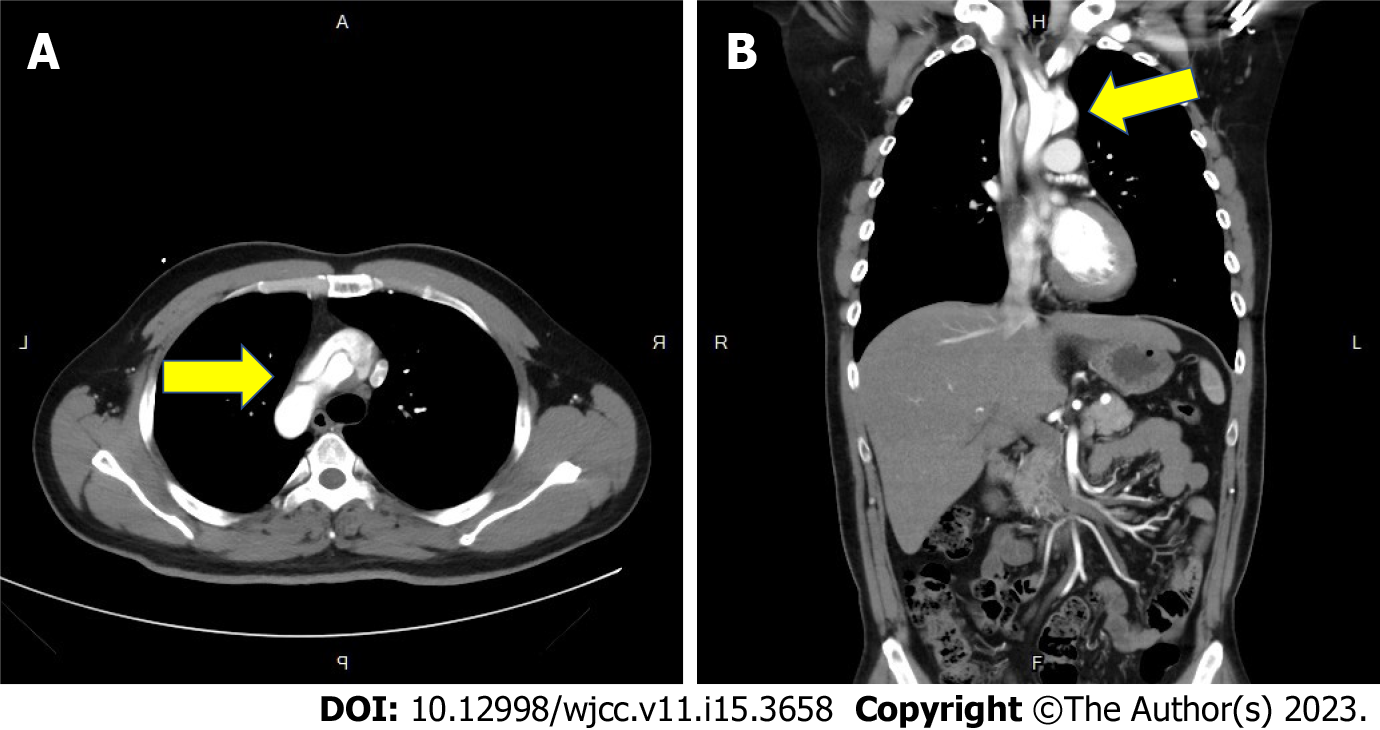
Figure 1 Type A aortic dissection with intimal flap involving the ascending aorta, the aortic arch and the common carotid arteries bilaterally.
A: The ascending aorta and the aortic arch (arrow); B: Arrow points to the mural thrombus in the false lumen.
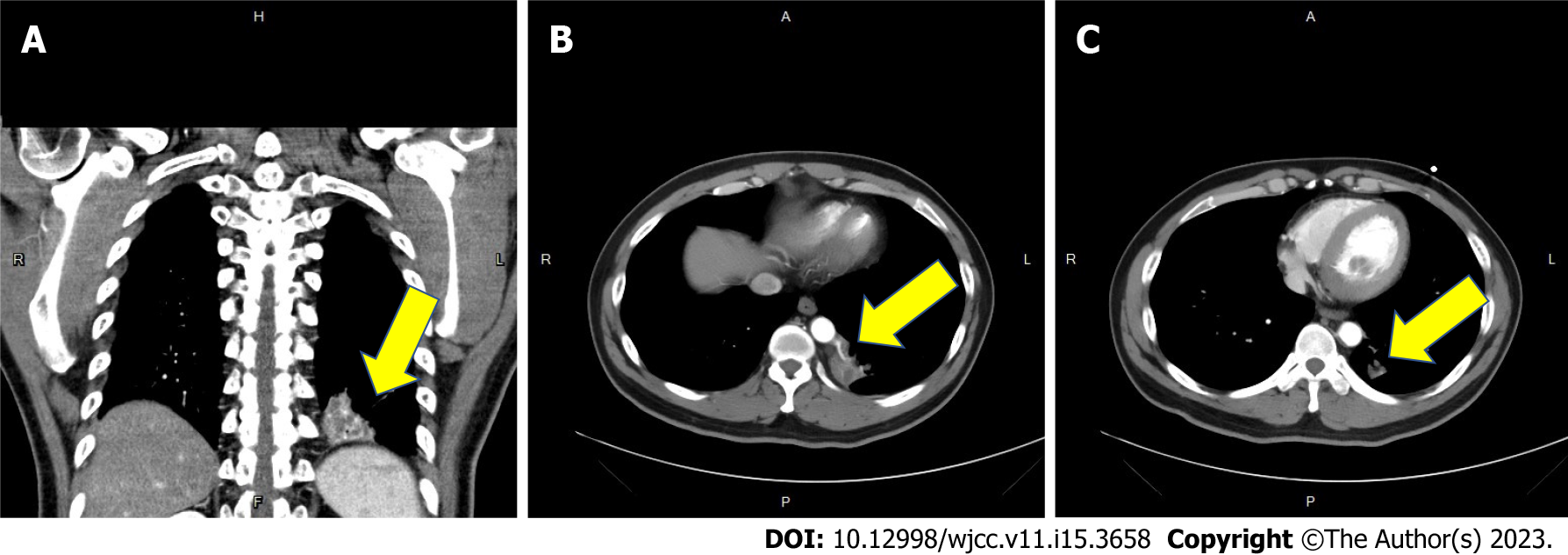
Figure 2 The contrast-enhanced computed tomography of the chest revealed a focal heterogeneous area with cystic change and patchy consolidation in the medial region of left lower lung with two aberrant arteries arising from the thoracic aorta supplying this area.
A: Left lower lung (arrow); B: Two aberrant arteries (arrow); C: Computed tomography angiography also revealed mild mural thickening and wall enhancement which may indicate mild vasculitis (arrow).
Figure 3 Video-assisted thoracoscopic surgery revealed that the two aberrant arteries originated from the thoracic aorta.
A: Two aberrant arteries (green arrow). Engorgement of the bronchus (yellow arrow) was also noticed. B: Gross specimen showed focal bronchial dilation that contained mucus (yellow arrow).
- Citation: Wang YJ, Chen YY, Lin GH. Relationship between intralobar pulmonary sequestration and type A aortic dissection: A case report. World J Clin Cases 2023; 11(15): 3658-3663
- URL: https://www.wjgnet.com/2307-8960/full/v11/i15/3658.htm
- DOI: https://dx.doi.org/10.12998/wjcc.v11.i15.3658


