©The Author(s) 2023.
World J Clin Cases. May 26, 2023; 11(15): 3592-3598
Published online May 26, 2023. doi: 10.12998/wjcc.v11.i15.3592
Published online May 26, 2023. doi: 10.12998/wjcc.v11.i15.3592
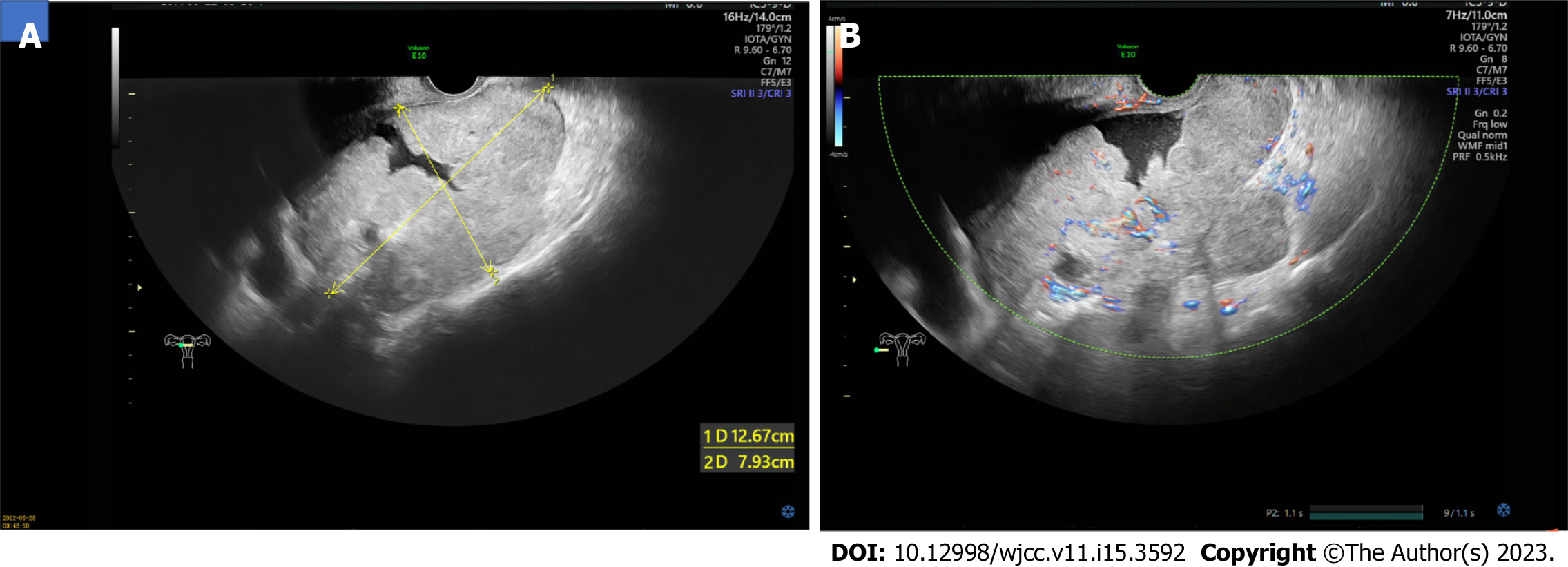
Figure 1 Transvaginal ultrasound images of the mass.
A: One mass of heterogeneous weak echo about 13 cm × 8 cm × 12 cm in size posterior to the uterus was detected; B: Inside the mass and surrounds were richly vascularized on color Doppler examination.
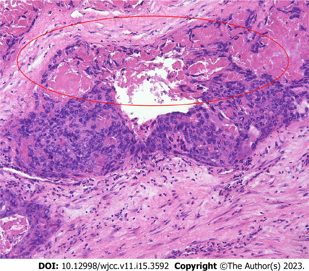
Figure 2 Intraoperative frozen pathological findings show choriocarcinoma with the presence of hemorrhage and necrosis within the tumor tissue and absence of ovarian tissue.
Haematoxylin and eosin staining (× 100).
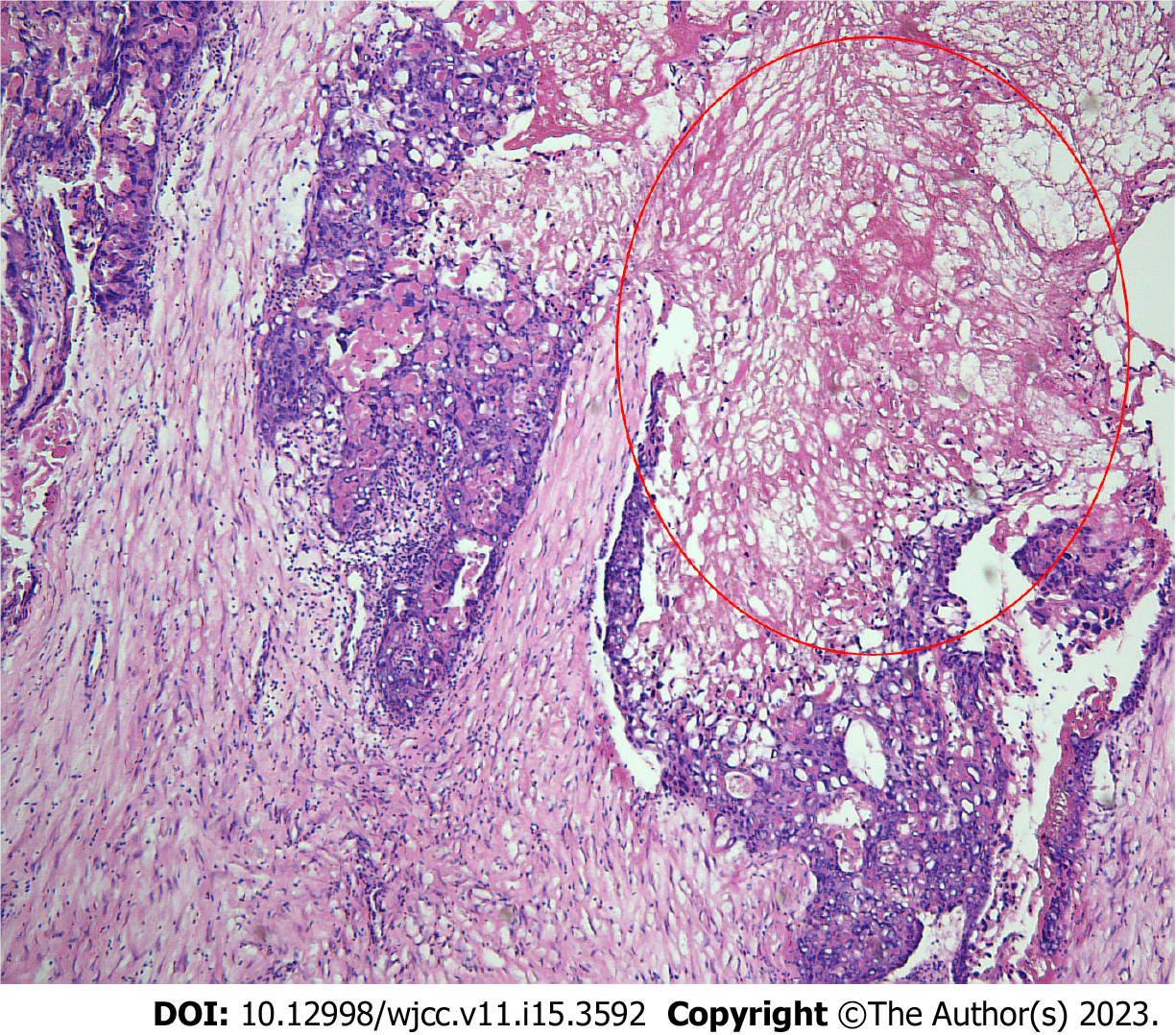
Figure 3 Postoperative microscopic appearance of the mass shows a pure choriocarcinoma with widespread necrosis.
Haematoxylin and eosin staining (× 40).
- Citation: Dai GL, Tang FR, Wang DQ. Primary ovarian choriocarcinoma occurring in a postmenopausal woman: A case report. World J Clin Cases 2023; 11(15): 3592-3598
- URL: https://www.wjgnet.com/2307-8960/full/v11/i15/3592.htm
- DOI: https://dx.doi.org/10.12998/wjcc.v11.i15.3592


