Copyright
©The Author(s) 2023.
World J Clin Cases. May 16, 2023; 11(14): 3356-3361
Published online May 16, 2023. doi: 10.12998/wjcc.v11.i14.3356
Published online May 16, 2023. doi: 10.12998/wjcc.v11.i14.3356
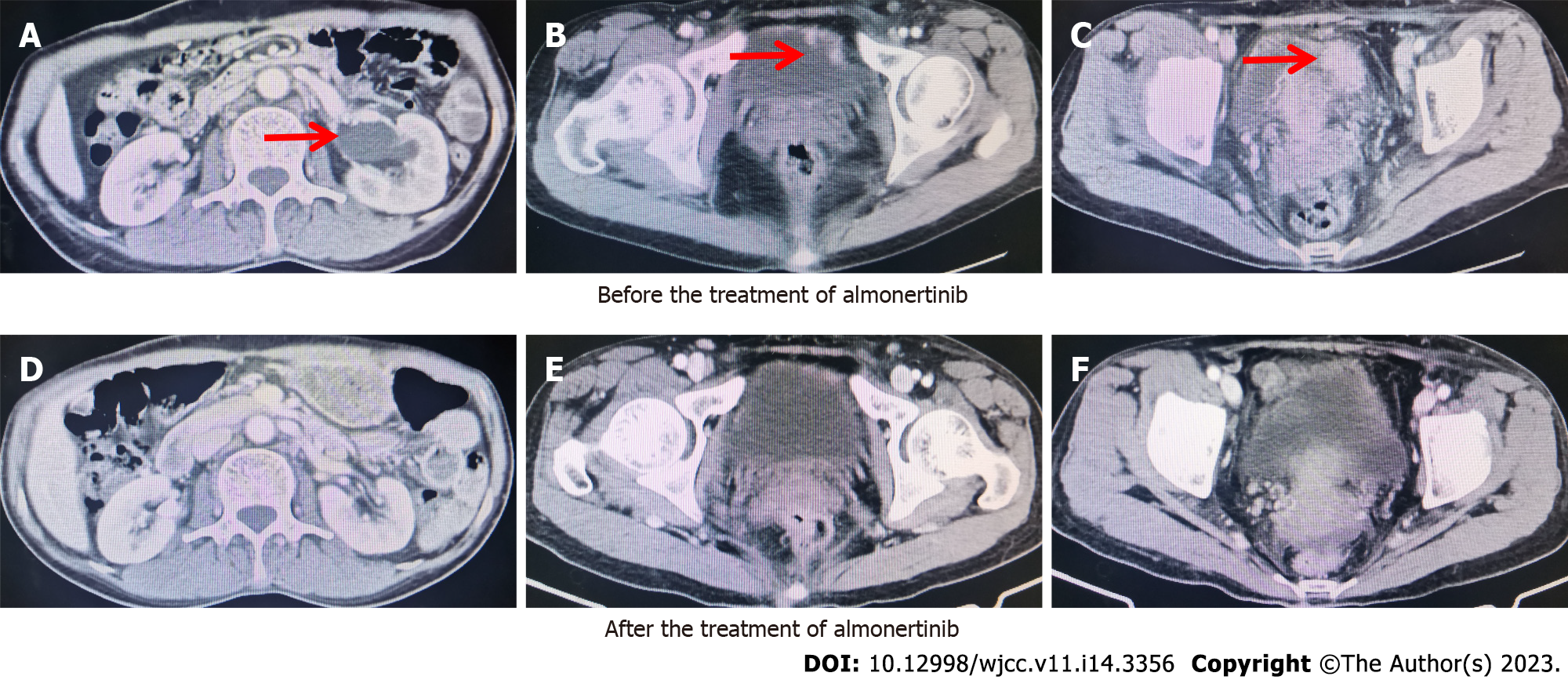
Figure 1 Computed tomography images recorded tumor regression of bladder metastasis after almonertinib therapy.
A-C: Before treatment; D-F: After treatment.
Figure 2 Cystoscopy analysis revealed multiple solid lesions on the anterior and bottom walls of the bladder.
A: Solid lesion on the anterior wall; B and C: Solid lesion on the bottom wall.
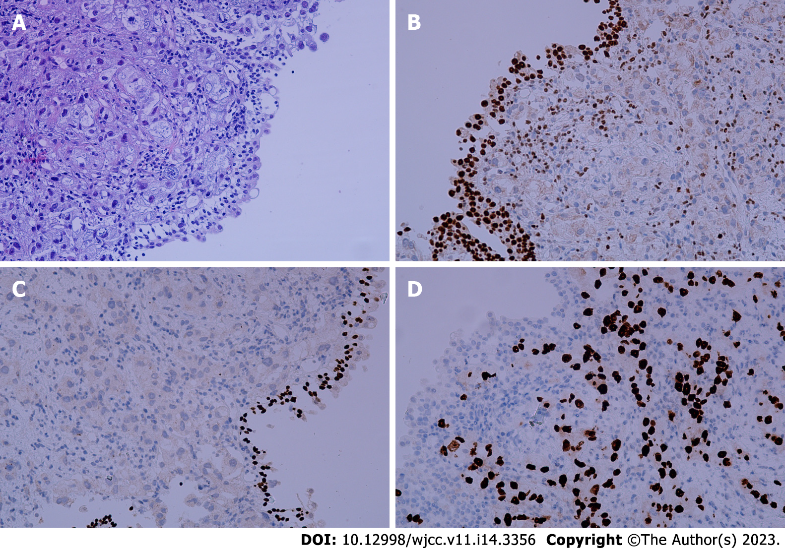
Figure 3 Histopathological findings in metastatic bladder sample.
A: Hematoxylin and eosin stained sections; B Negative staining for GATA3; C: Negative staining for P40; D: Positive staining for thyroid transcription factor 1.
- Citation: Jin CB, Yang L. Bladder metastasis from epidermal growth factor receptor mutant lung cancer: A case report. World J Clin Cases 2023; 11(14): 3356-3361
- URL: https://www.wjgnet.com/2307-8960/full/v11/i14/3356.htm
- DOI: https://dx.doi.org/10.12998/wjcc.v11.i14.3356


