Copyright
©The Author(s) 2023.
World J Clin Cases. May 16, 2023; 11(14): 3204-3210
Published online May 16, 2023. doi: 10.12998/wjcc.v11.i14.3204
Published online May 16, 2023. doi: 10.12998/wjcc.v11.i14.3204
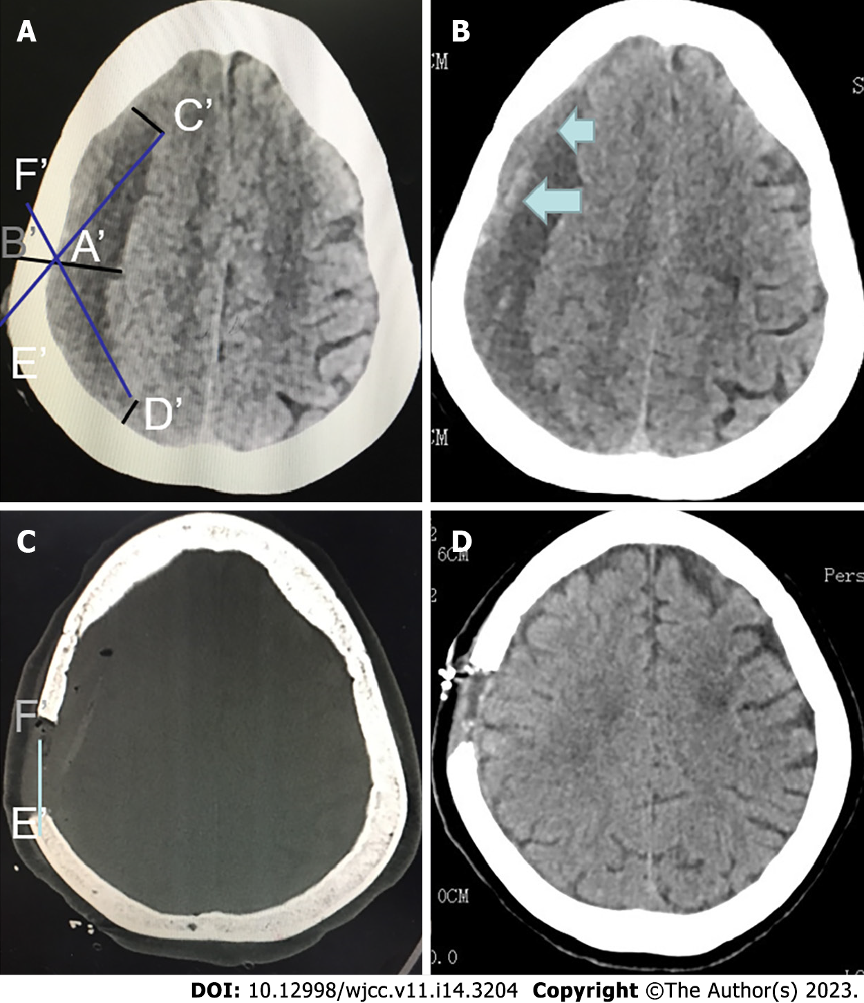
Figure 1 Head computed tomography imaging and surgical design.
A: On the thickest layer of the hematoma cavity in the computed tomography (CT) image, make a straight line perpendicular to brain surface, which cross the skull at point A' at the inner side and point B' at the outer side. At the edge of the hematoma at the layer, find points C' and D' with distance to A' and B' equal to the thickness of the hematoma, respectively. Connect and extend C'A' and D'A', which intersect at points E' and F' with the surface of the skull, respectively; B: Axial CT scan showing septated right chronic subdural hematoma with two clear compartments. Small blue arrows indicate the membrane to be opened; C: Axial CT scan showing the range of bone window; and D: Axial CT scan showing the postoperative intracranial conditions. CT: Computed tomography.
Figure 2 Surgical incision and intraoperative image.
A: Preoperative planning of incisions; B: Postoperative planning of incisions; and C: Endoscopic view of the subdural space identifying the membrane with its rich microvascularization and organized blood clot.
- Citation: Wang XJ, Yin YH, Zhang LY, Wang ZF, Sun C, Cui ZM. Positioning and design by computed tomography imaging in neuroendoscopic surgery of patients with chronic subdural hematoma. World J Clin Cases 2023; 11(14): 3204-3210
- URL: https://www.wjgnet.com/2307-8960/full/v11/i14/3204.htm
- DOI: https://dx.doi.org/10.12998/wjcc.v11.i14.3204


