Copyright
©The Author(s) 2023.
World J Clin Cases. Apr 16, 2023; 11(11): 2482-2488
Published online Apr 16, 2023. doi: 10.12998/wjcc.v11.i11.2482
Published online Apr 16, 2023. doi: 10.12998/wjcc.v11.i11.2482
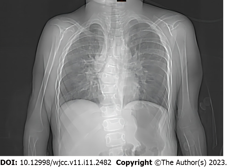
Figure 1 Chest radiography demonstrated pneumonia, scoliosis, and right deviation of the trachea.
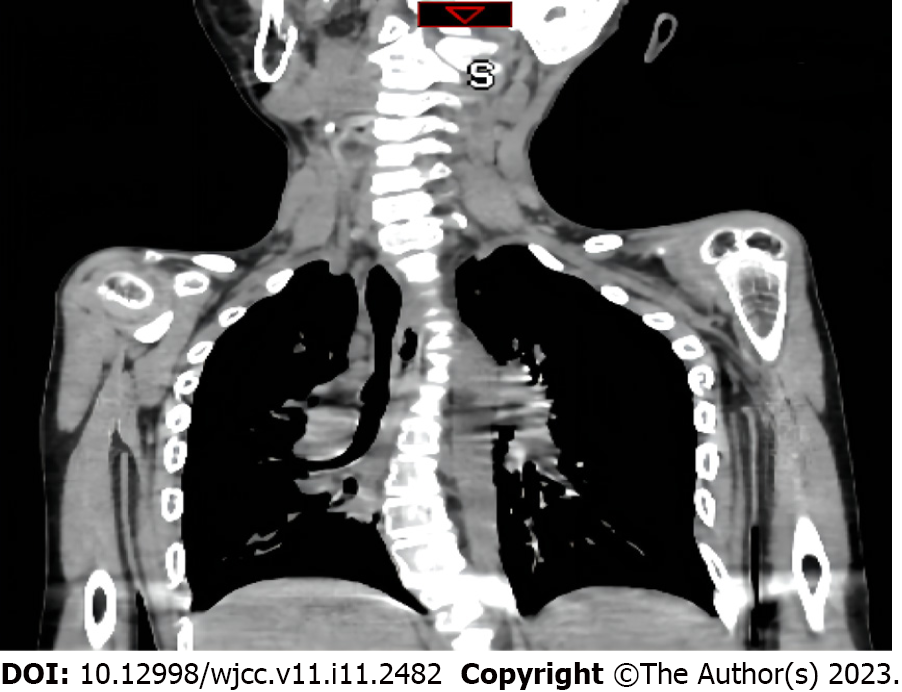
Figure 2 Computed tomography scans revealed scoliosis, osteoporosis of the spine, and thoracic and tracheal malformation.
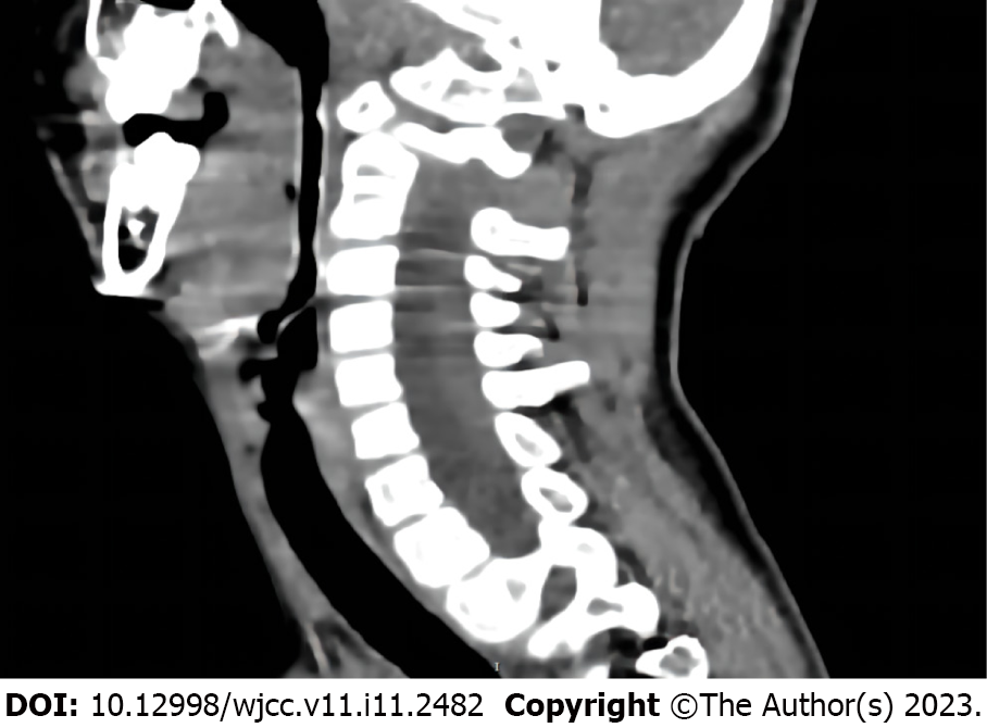
Figure 3 Lateral cervical spine computed tomography scans displayed laryngomalacia, malformations of the pharynx and cervical spine.
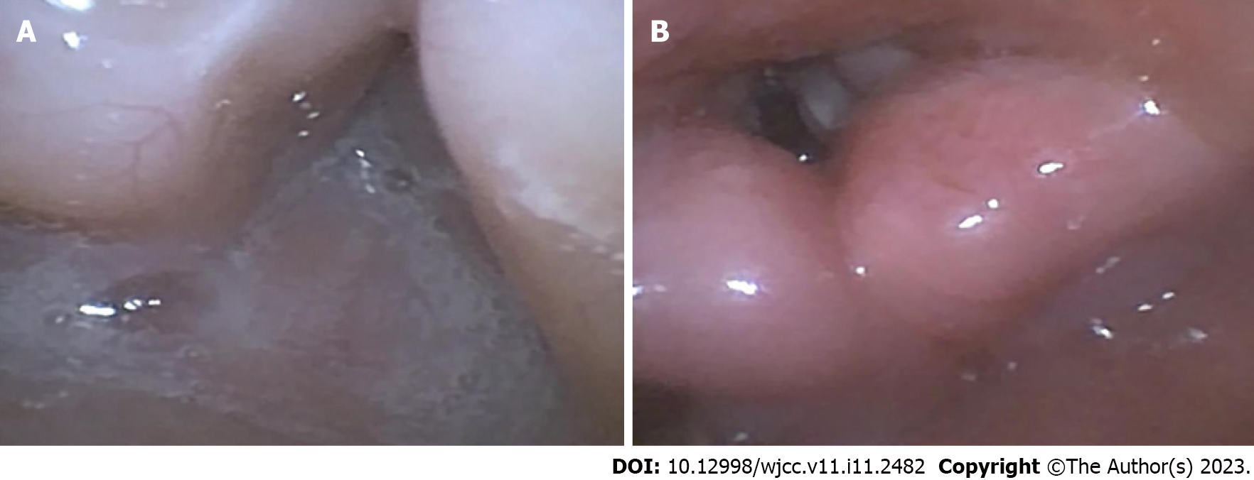
Figure 4 Fiberoptic bronchoscopy.
A: There were massive secretions in the nasopharynx; B: The glottis was clearly visible in the transoral approach.
- Citation: Chen JX, Shi XL, Liang CS, Ma XG, Xu L. Anesthesia management in a pediatric patient with complicatedly difficult airway: A case report. World J Clin Cases 2023; 11(11): 2482-2488
- URL: https://www.wjgnet.com/2307-8960/full/v11/i11/2482.htm
- DOI: https://dx.doi.org/10.12998/wjcc.v11.i11.2482


