Copyright
©The Author(s) 2023.
World J Clin Cases. Apr 16, 2023; 11(11): 2464-2473
Published online Apr 16, 2023. doi: 10.12998/wjcc.v11.i11.2464
Published online Apr 16, 2023. doi: 10.12998/wjcc.v11.i11.2464
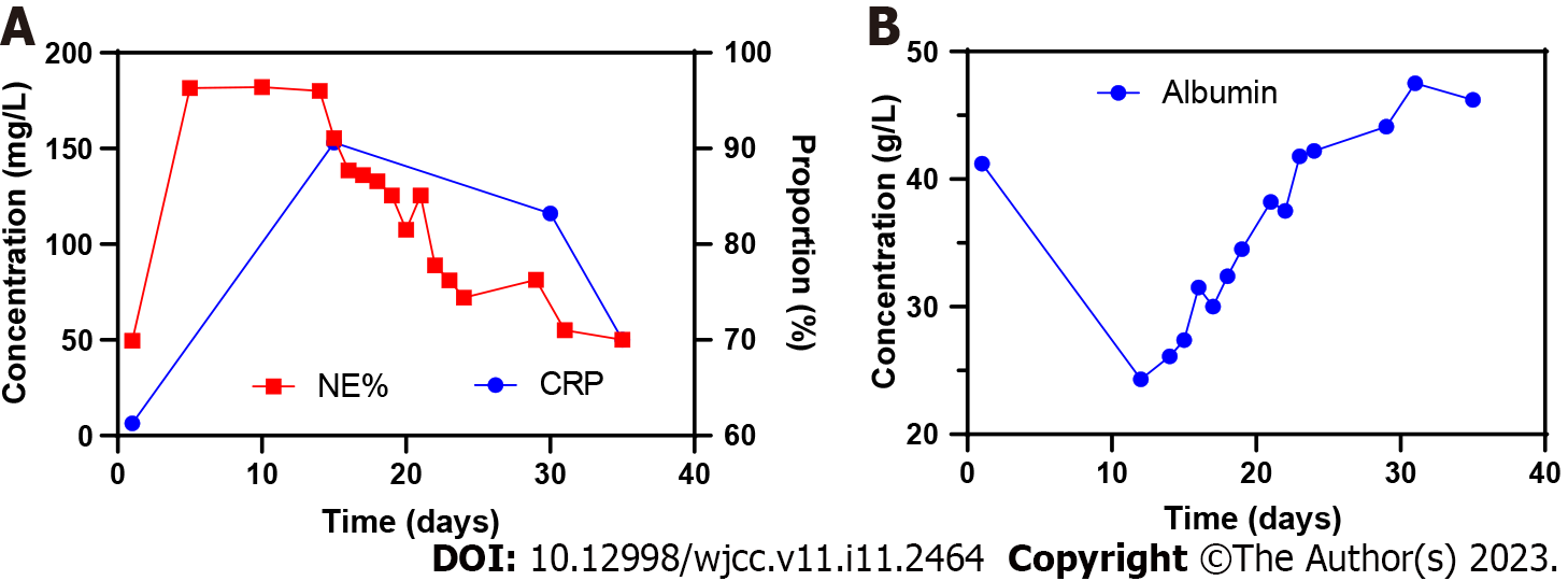
Figure 1 Changes in C-reactive protein level, neutrophils percentage, and albumin level during hospitalization of the first patient.
A: Changes in C-reactive protein level, neutrophils percentage; B: Changes in albumin level. CRP: C-reactive protein; NE%: Neutrophils percentage.
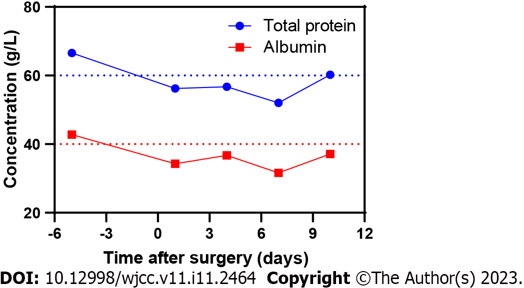
Figure 2 Nutritional status of the second patient during hospitalization.
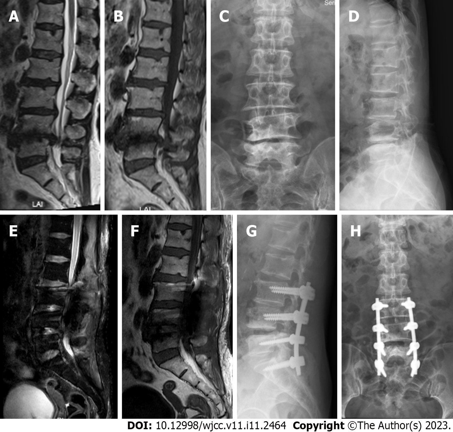
Figure 3 Magnetic resonance imaging and X-ray findings of the first patient.
A–D: Preoperative images showing lumbar disc herniation and lumbar spinal stenosis at L3–S1 level; E–H: Images at 1 wk after surgery showing the lack of clear signs of cerebrospinal fluid leakage or pseudocyst formation.
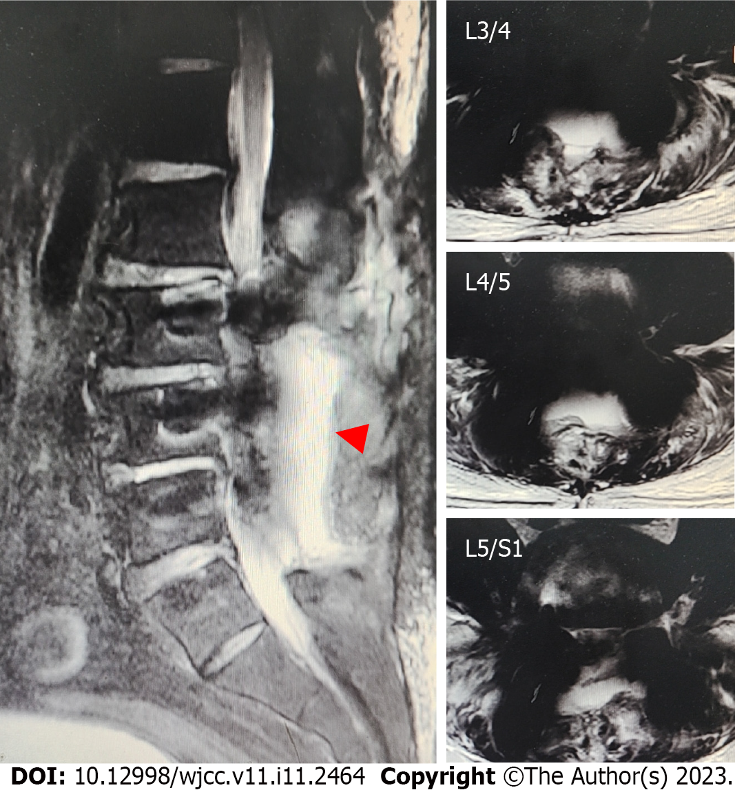
Figure 4 Magnetic resonance imaging findings of the second patient.
Red triangle: Exuded cerebrospinal fluid.
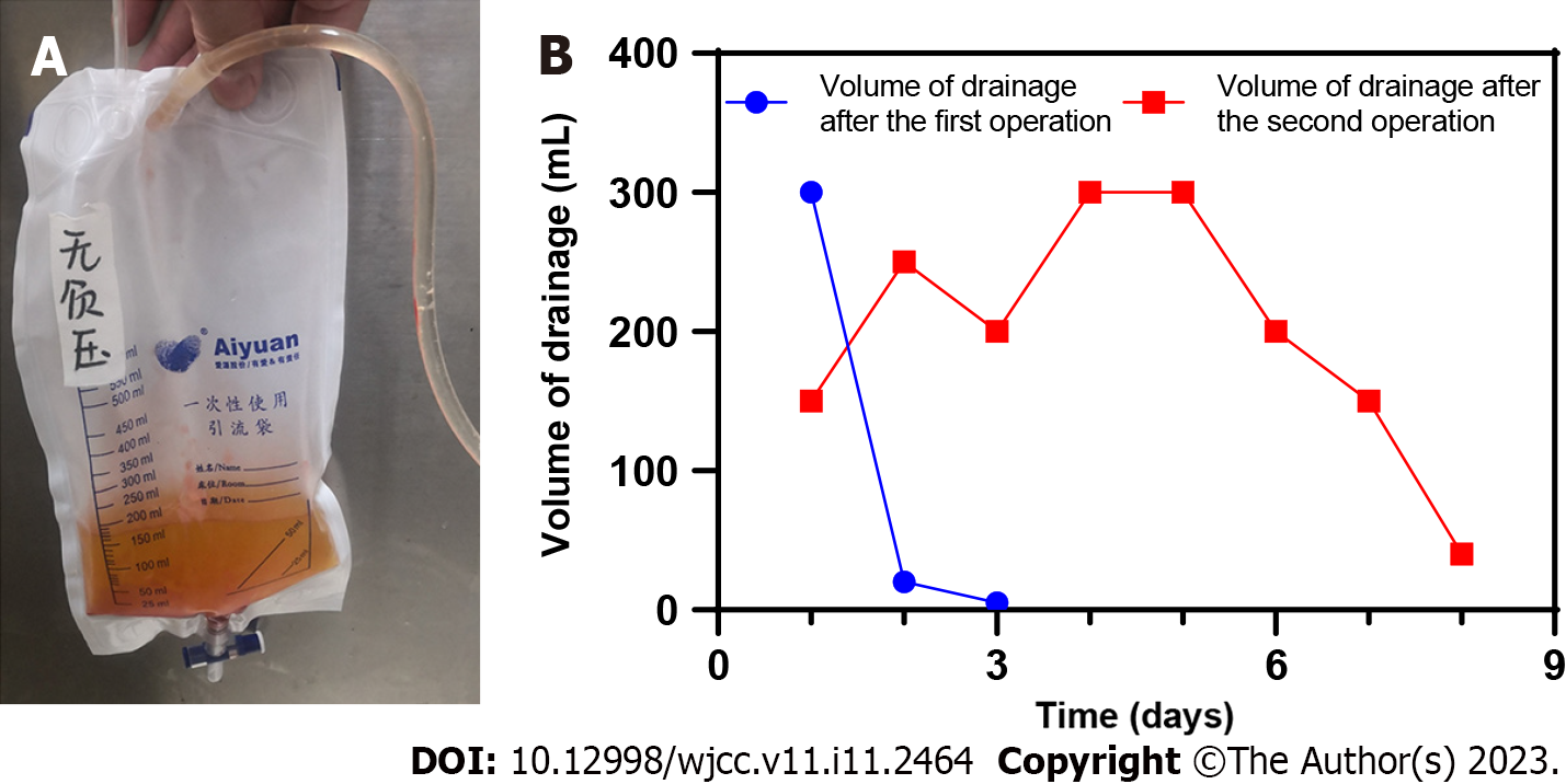
Figure 5 Postoperative drainage of the first patient.
A: Drainage fluid after the third operation. The drainage fluid is light red, which is considered as cerebrospinal fluid; B: Changes of postoperative drainage volume.
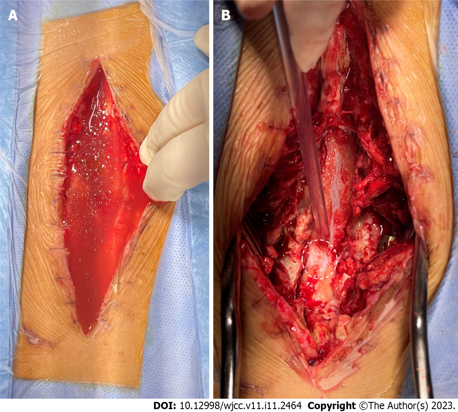
Figure 6 Digital photos of intraoperative exploration of the second patient.
A: A large amount of liquid accumulated under the skin; B: Dural defect found during operation. White dotted circle: The size of dura defect.
- Citation: Xu C, Dong RP, Cheng XL, Zhao JW. Late presentation of dural tears: Two case reports and review of literature. World J Clin Cases 2023; 11(11): 2464-2473
- URL: https://www.wjgnet.com/2307-8960/full/v11/i11/2464.htm
- DOI: https://dx.doi.org/10.12998/wjcc.v11.i11.2464


