©The Author(s) 2023.
World J Clin Cases. Apr 6, 2023; 11(10): 2343-2348
Published online Apr 6, 2023. doi: 10.12998/wjcc.v11.i10.2343
Published online Apr 6, 2023. doi: 10.12998/wjcc.v11.i10.2343
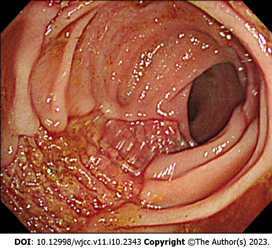
Figure 1 Sigmoidoscopy image.
Sigmoidoscopy image shows presenting a longitudinal ulcer from the anal verge (AV) to 12 cm above the AV.
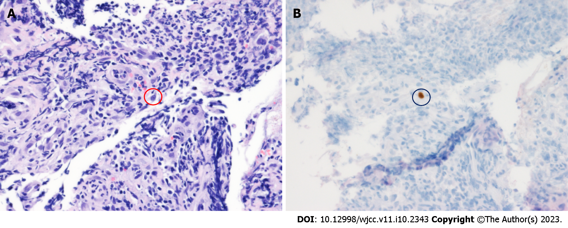
Figure 2 Pathology findings.
A: Red circle shows a cell with a basophilic intranuclear inclusion body surrounded by a clear halo (hematoxylin and eosin stain, 400 ×); B: Black circle shows positive immunohistochemistry for cytomegalovirus (immunohistochemical stain, 400 ×).
- Citation: Kim JH, Kim HS, Jeong HW. Coexisting cytomegalovirus colitis in an immunocompetent patient with Clostridioides difficile colitis: A case report. World J Clin Cases 2023; 11(10): 2343-2348
- URL: https://www.wjgnet.com/2307-8960/full/v11/i10/2343.htm
- DOI: https://dx.doi.org/10.12998/wjcc.v11.i10.2343


