©The Author(s) 2023.
World J Clin Cases. Apr 6, 2023; 11(10): 2336-2342
Published online Apr 6, 2023. doi: 10.12998/wjcc.v11.i10.2336
Published online Apr 6, 2023. doi: 10.12998/wjcc.v11.i10.2336
Figure 1 Café-au-lait patches and cutaneous nodular masses.
A: Multiple cutaneous nodular masses on the head and face; B: Multiple café-au-lait patches and cutaneous nodular masses on the back.
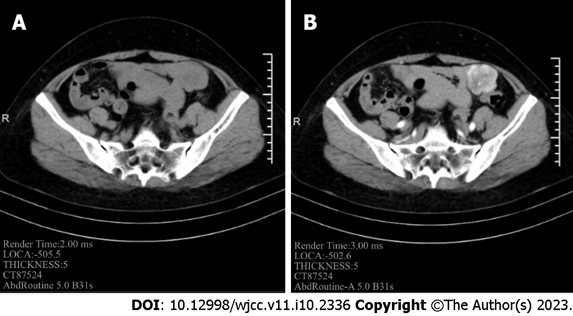
Figure 2 Abdominal computed tomography.
A: A cystic solid mass with an oblique diameter of approximately 44 mm is seen in the left pelvis; B: The solid component exhibits arterial enhancement.
Figure 3 Intraoperative view.
A: Multiple masses in the jejunal wall demonstrated clear borders; B: A 4.5 cm diameter tumor in the jejunum 20 cm from the Trietz ligament; C: Large tumor with a grayish white, nodular, cystic surface; D: Small tumor with an oval shape and solid nodules.
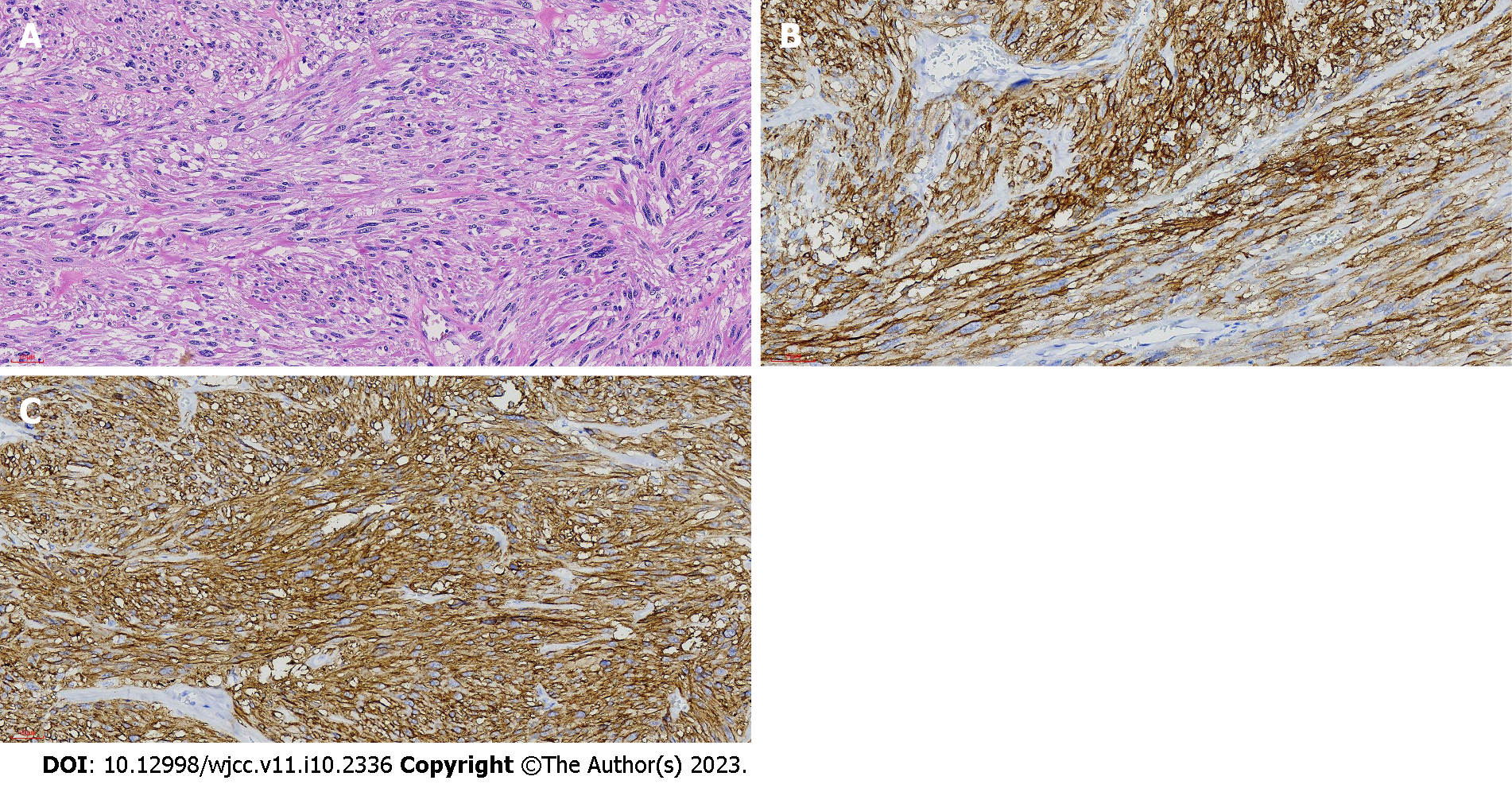
Figure 4 Postoperative pathology and immunohistochemical results.
A: Diffuse spindle cells with rod-shaped nuclei (hematoxylin–eosin, × 200); B: Diffusely strong expression of CD117 (× 200); C: Diffusely strong expression of DOG-1 (× 200).
- Citation: Yao MQ, Jiang YP, Yi BH, Yang Y, Sun DZ, Fan JX. Neurofibromatosis type 1 with multiple gastrointestinal stromal tumors: A case report. World J Clin Cases 2023; 11(10): 2336-2342
- URL: https://www.wjgnet.com/2307-8960/full/v11/i10/2336.htm
- DOI: https://dx.doi.org/10.12998/wjcc.v11.i10.2336


