Copyright
©The Author(s) 2023.
World J Clin Cases. Apr 6, 2023; 11(10): 2282-2289
Published online Apr 6, 2023. doi: 10.12998/wjcc.v11.i10.2282
Published online Apr 6, 2023. doi: 10.12998/wjcc.v11.i10.2282
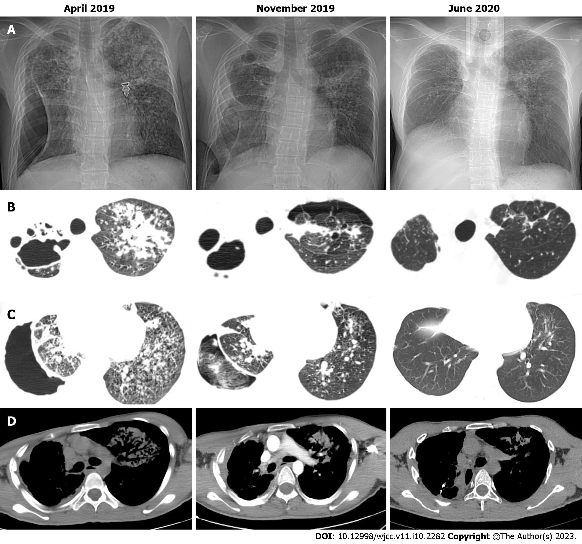
Figure 1 Chest computed tomography scans of patient.
A: At admission and before surgery show bilateral pulmonary infection and complete compression and atelectasis of the right lung. Postoperative chest computed tomography (CT) demonstrates right lung expansion; B: Chest CT scan showing the destruction of the right upper lung; C: Both the presence of pus in the pleural cavity and iodine gauze in the emphysematous pleural space are observed. The residual cavity is eliminated after surgery; D: The coarctation of the right intercostal space is noted on chest CT. The patient’s right chest wall deformity has almost resolved, 6 months after the operation.
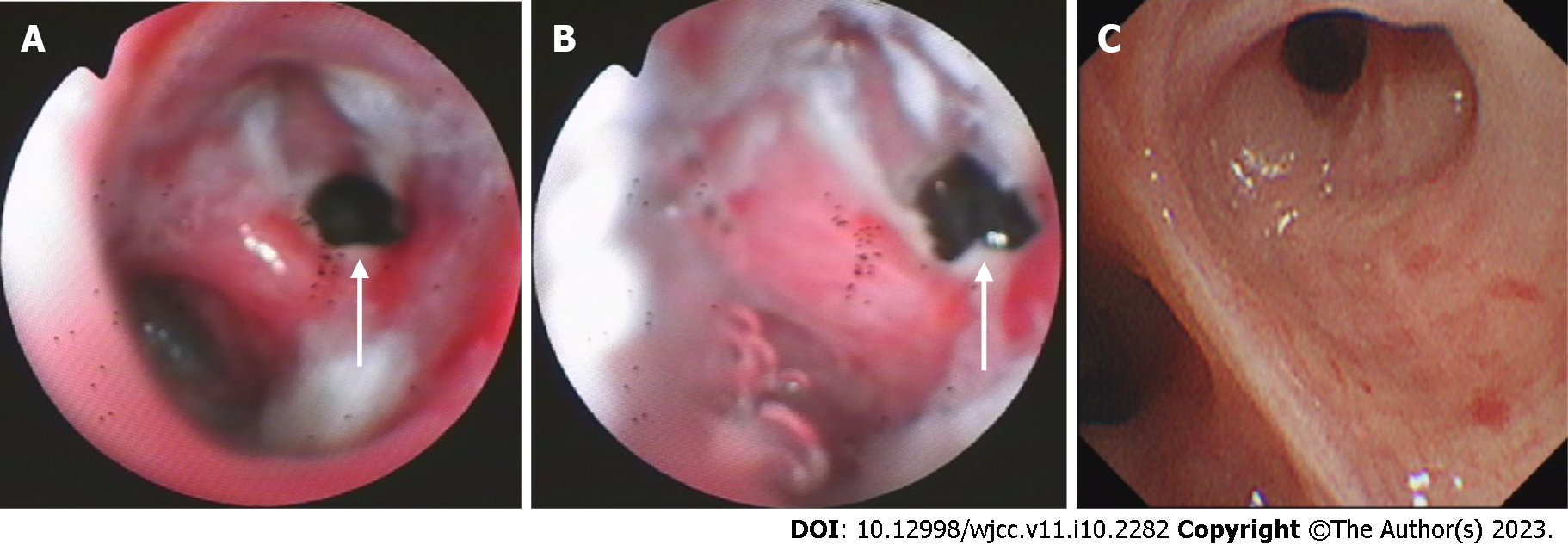
Figure 2 Bronchoscopic view of the right upper bronchus.
A: Bronchoscopy at admission shows a 5 mm fistula (white arrows) of the right upper bronchus, along with erythema, erosion, and hyperplasia of the apical bronchus, and a small amount of necrotic material; B: Bronchoscopy is performed 1 mo after the anti-tuberculosis treatment; C: Bronchoscopy is performed 6 mo after the anti-tuberculosis treatment.
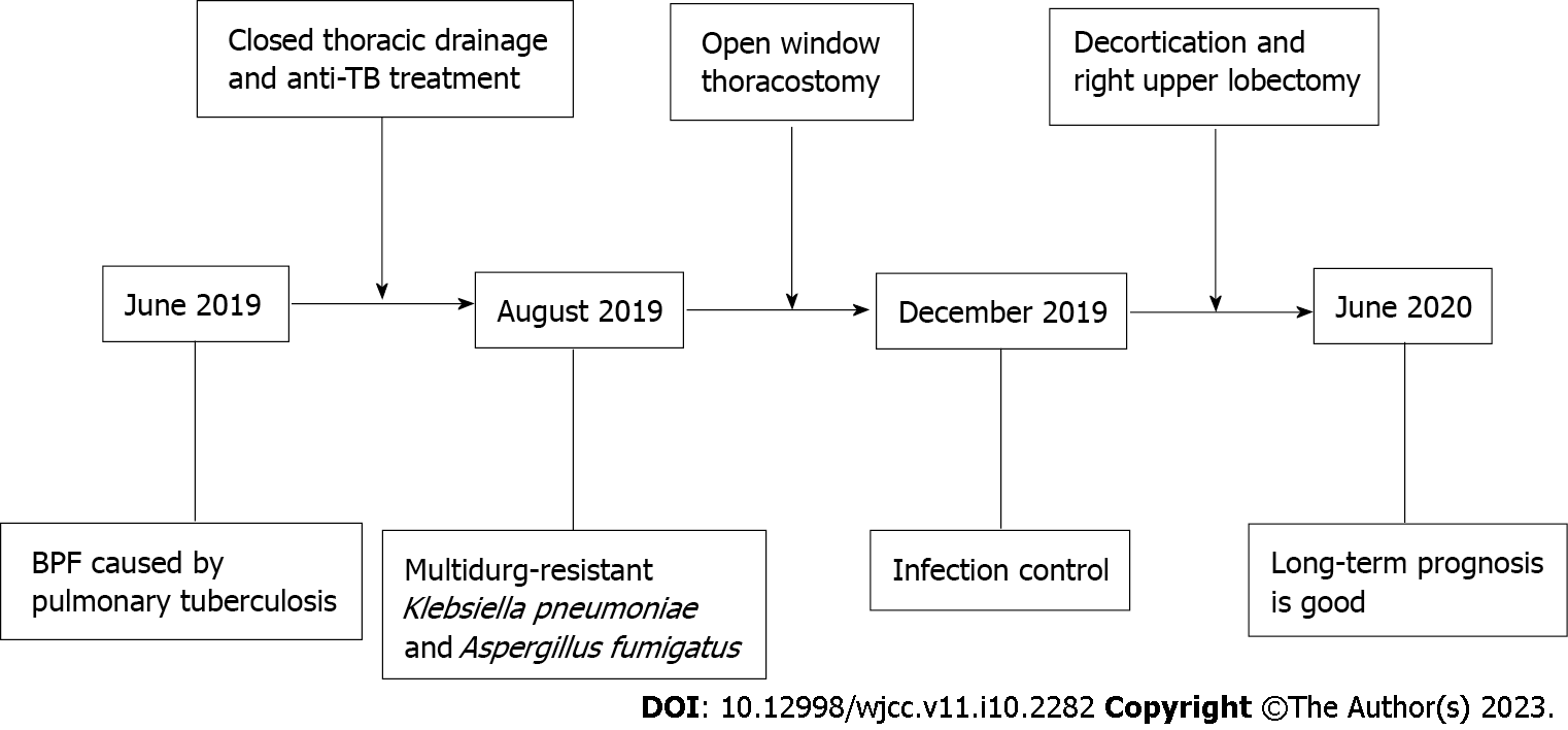
Figure 3 The timeline of treatment and procedures.
TB: Tuberculosis; BPF: Bronchopleural fistula.
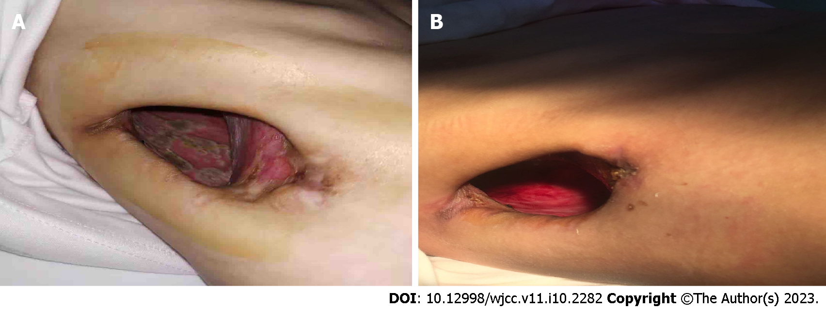
Figure 4 Intraoperative and postoperative findings.
A: Open-window thoracostomy was performed on the patient; intraoperatively, mold was noted on the pleural surface; B: No effusions were noted in the pleural cavity and no lesions were seen on the pleural surface.
- Citation: Shen L, Jiang YH, Dai XY. Successful surgical treatment of bronchopleural fistula caused by severe pulmonary tuberculosis: A case report and review of literature. World J Clin Cases 2023; 11(10): 2282-2289
- URL: https://www.wjgnet.com/2307-8960/full/v11/i10/2282.htm
- DOI: https://dx.doi.org/10.12998/wjcc.v11.i10.2282


