Copyright
©The Author(s) 2022.
World J Clin Cases. Mar 16, 2022; 10(8): 2559-2568
Published online Mar 16, 2022. doi: 10.12998/wjcc.v10.i8.2559
Published online Mar 16, 2022. doi: 10.12998/wjcc.v10.i8.2559
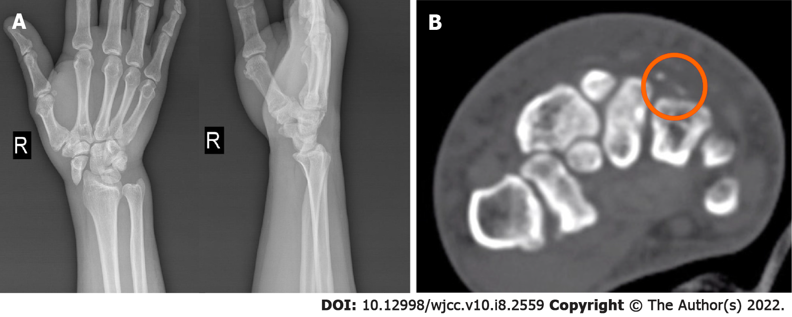
Figure 1 Initial radiographs demonstrated primary complex partial radial dislocation of the scaphoid; The computed tomography scans demonstrated the chip fractures of the capitate and hamate.
A: Posteroanterior and lateral; B: Computed tomography scans.
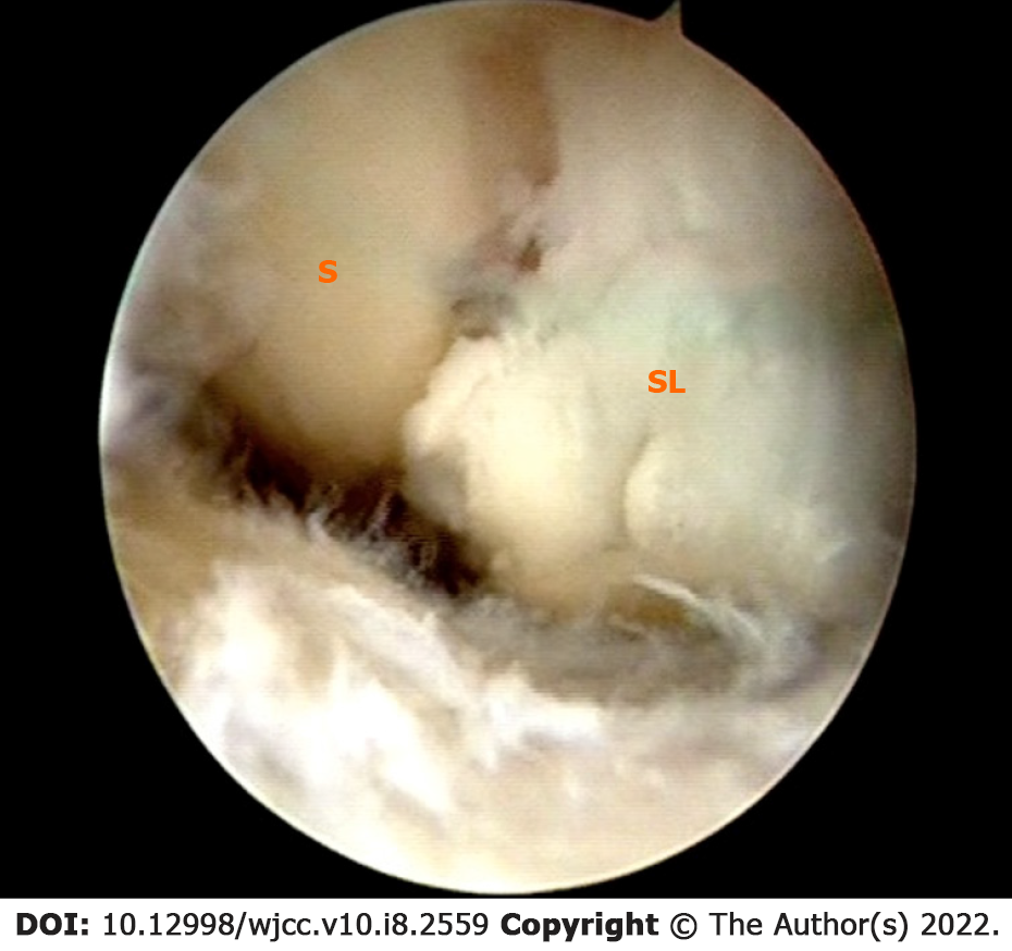
Figure 2 Arthroscopy confirmed complete disruption of the scapholunate and radioscaphocapitate ligaments, and scapholunate diastasis was Geissler grade IV.
S: Scaphoid; SL: Scapholunate ligament.
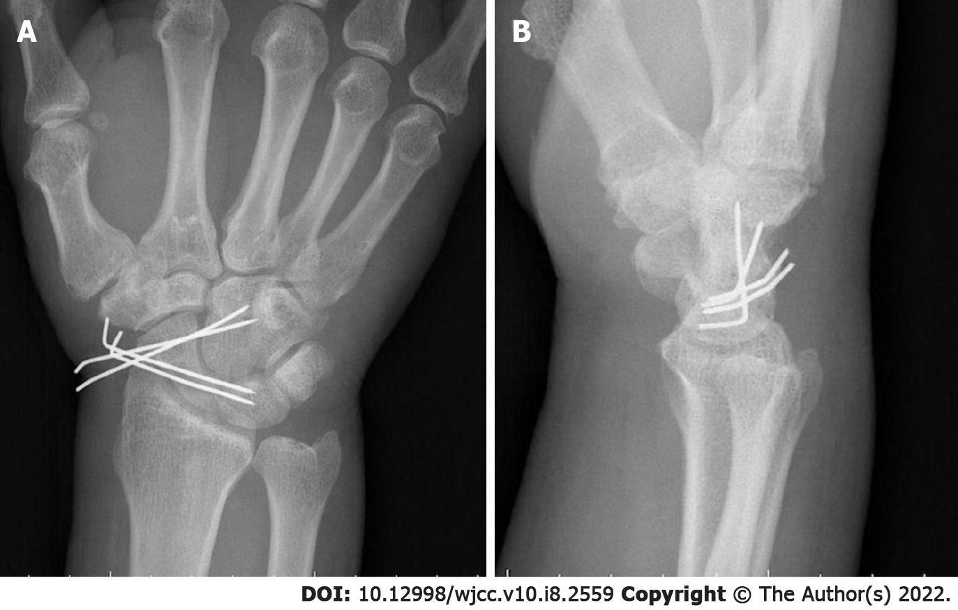
Figure 3 Postoperative radiographs showed internal fixation with K-wires.
A: Posteroanterior; B: Lateral.
Figure 4 At the 6-mo follow-up, the action of all sides of the carpal bone.
A: Normal flexion; B: Slight restriction of extension; C: Supination; D: Pronation. These four movements were observed compared to the contralateral side.
Figure 5 At the 6-mo follow-up, radiographs revealed neither avascular necrosis of the proximal pole of the scaphoid nor scapholunate diastasis.
A: Posteroanterior; B: Lateral.
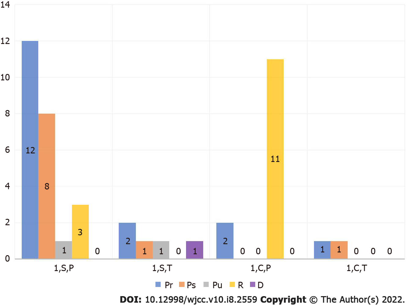
Figure 6 Classifications of isolated scaphoid dislocation.
1: Primary; C: Complex; D: Dorsal; P: Partial; Pr: Palmar radial; Ps: Palmar straight; Pu: Palmar ulnar[2]; R: Pure radial; S: Simple; T: Total.
- Citation: Liu SD, Yin BS, Han F, Jiang HJ, Qu W. Isolated scaphoid dislocation: A case report and review of literature. World J Clin Cases 2022; 10(8): 2559-2568
- URL: https://www.wjgnet.com/2307-8960/full/v10/i8/2559.htm
- DOI: https://dx.doi.org/10.12998/wjcc.v10.i8.2559


