Copyright
©The Author(s) 2022.
World J Clin Cases. Feb 6, 2022; 10(4): 1326-1332
Published online Feb 6, 2022. doi: 10.12998/wjcc.v10.i4.1326
Published online Feb 6, 2022. doi: 10.12998/wjcc.v10.i4.1326
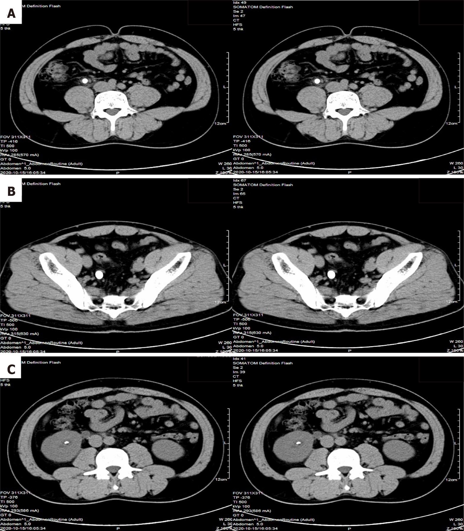
Figure 1 Abdominal computed tomography scans.
A: One stone in the right upper ureter; B: Multiple stones in the right lower ureter; C: Other stones in the right kidney.
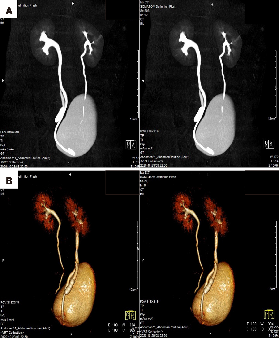
Figure 2 Computed tomography urography and three-dimensional reconstructions.
A: Computed tomography urography; B: Three-dimensional reconstructions. The middle of the right ureter was divided into two branches, suggesting duplicated ureteral malformation with an ectopic ureter.
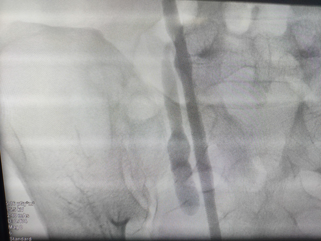
Figure 3
Intraoperative retrograde ureterography showed that the ectopic ureter was visible.
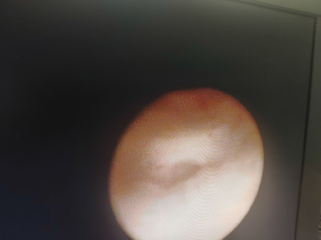
Figure 4
Duplicated ureteral bifurcation could be seen by endoscopy.
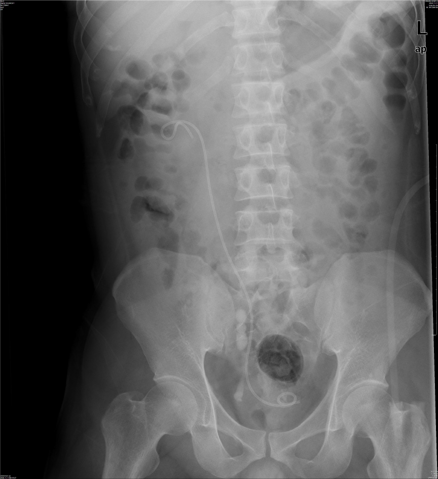
Figure 5
KUB after the operation showed that stones in the right upper ureter and renal pelvis had disappeared, a double J tube was placed, and multiple stones in the ectopic ureter were visible.
- Citation: Ye WX, Ren LG, Chen L. Inverted Y ureteral duplication with an ectopic ureter and multiple urinary calculi: A case report. World J Clin Cases 2022; 10(4): 1326-1332
- URL: https://www.wjgnet.com/2307-8960/full/v10/i4/1326.htm
- DOI: https://dx.doi.org/10.12998/wjcc.v10.i4.1326


