Copyright
©The Author(s) 2022.
World J Clin Cases. Dec 26, 2022; 10(36): 13458-13466
Published online Dec 26, 2022. doi: 10.12998/wjcc.v10.i36.13458
Published online Dec 26, 2022. doi: 10.12998/wjcc.v10.i36.13458
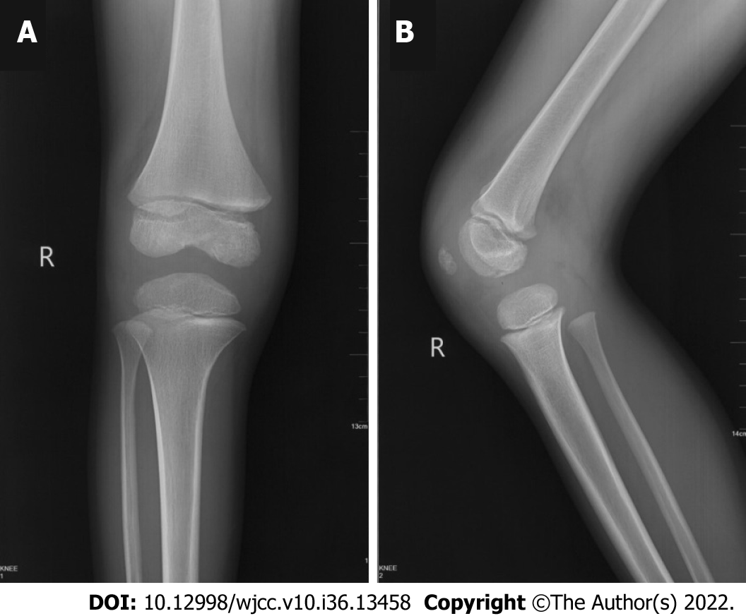
Figure 1 X-ray before surgery shows a stable fracture in the medial femoral condyle with no displacement.
A: Anterior-posterior view of X-ray; B: Lateral view of X-ray.
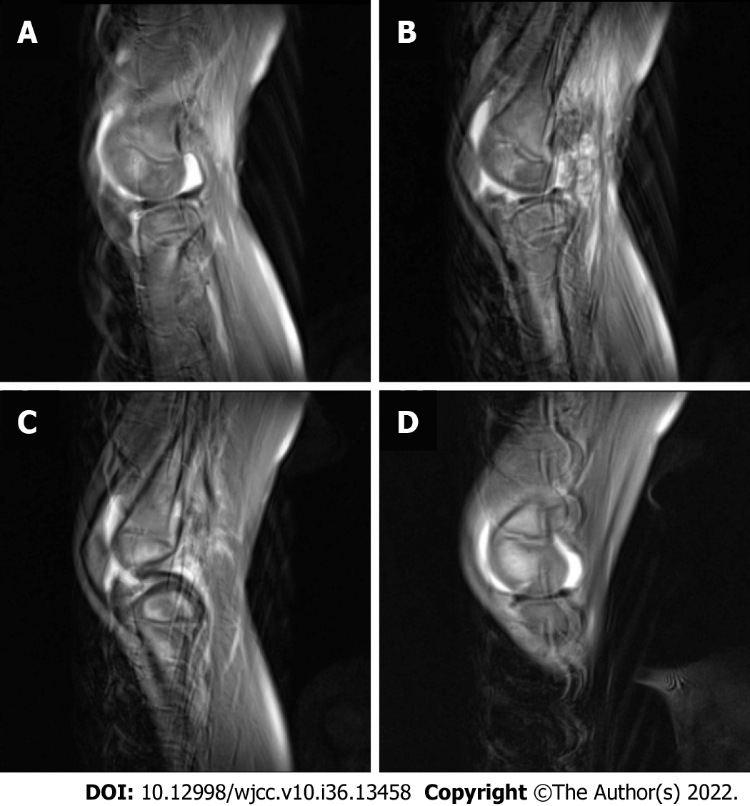
Figure 2 Magnetic resonance imaging before surgery indicated no severe displacement.
A: Sagittal slice of lateral condyle of femur; B: Most lateral slice of femoral intercondylar notch; C: Most medial slice of femoral intercondylar notch; D: Sagittal slice of medial condyle of femur.
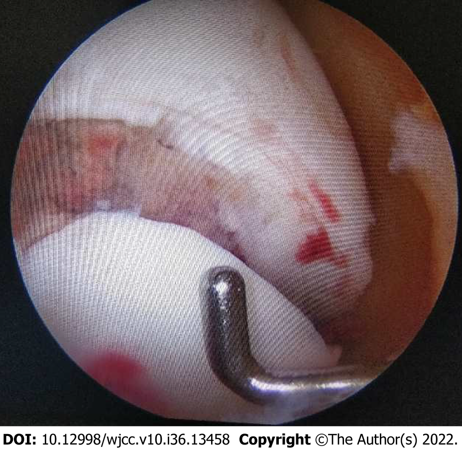
Figure 3
Arthroscopy showed obvious fracture displacement of the cartilage.
Figure 4
The operation was completed using the medial parapatellar approach.
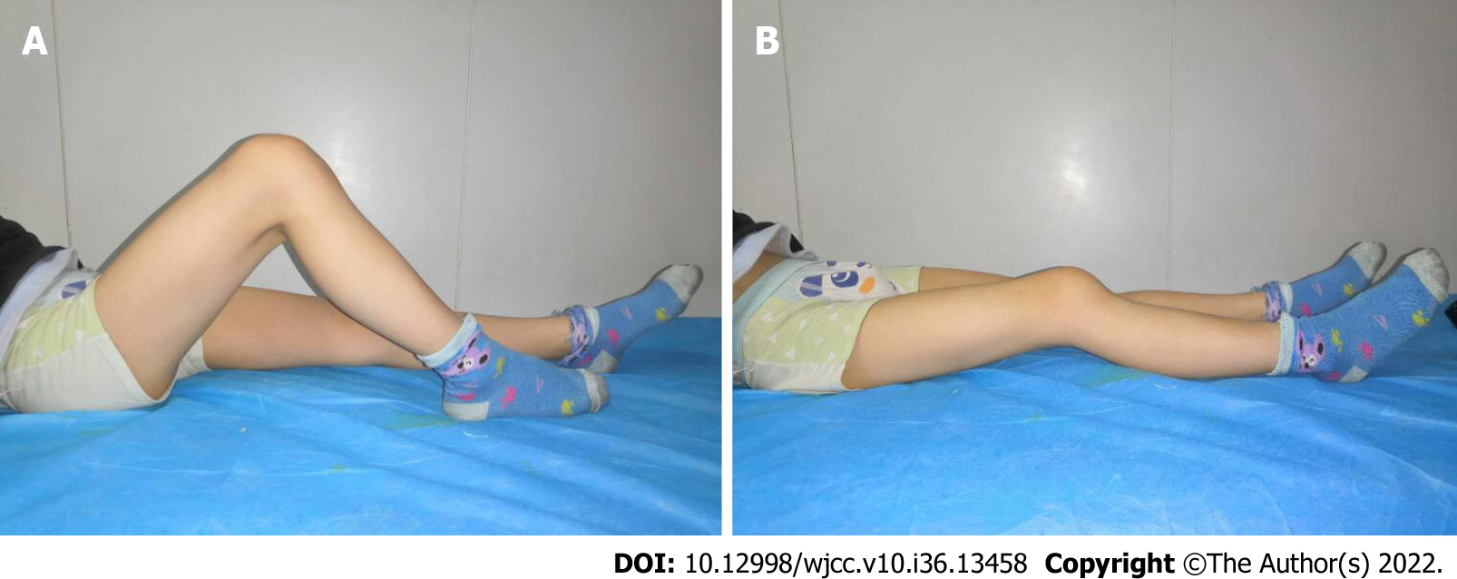
Figure 5 Six months after surgery, the range of motion of the knee joint reached 5°-100°.
A: Maximum flexion position; B: Maximum extension position.
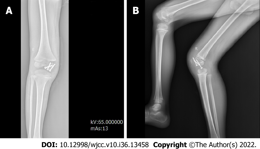
Figure 6 Plain radiographs showed a well-healed fracture with no evidence of collapse of the femoral condyle.
A: Anterior-posterior view of X-ray; B: Lateral view of X-ray.
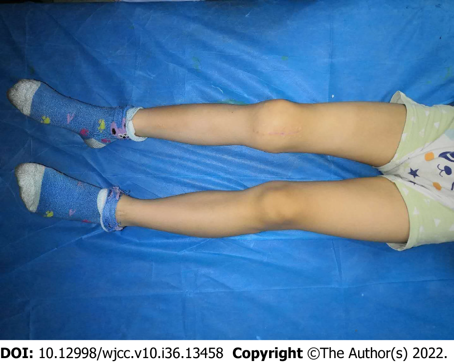
Figure 7 At the final follow-up of 40 months, the patient had full range of motion.
A: Maximum flexion position; B: Maximum extension position
- Citation: Chen ZH, Wang HF, Wang HY, Li F, Bai XF, Ni JL, Shi ZB. Hoffa's fracture in a five-year-old child diagnosed and treated with the assistance of arthroscopy: A case report. World J Clin Cases 2022; 10(36): 13458-13466
- URL: https://www.wjgnet.com/2307-8960/full/v10/i36/13458.htm
- DOI: https://dx.doi.org/10.12998/wjcc.v10.i36.13458


