Copyright
©The Author(s) 2022.
World J Clin Cases. Dec 26, 2022; 10(36): 13373-13380
Published online Dec 26, 2022. doi: 10.12998/wjcc.v10.i36.13373
Published online Dec 26, 2022. doi: 10.12998/wjcc.v10.i36.13373
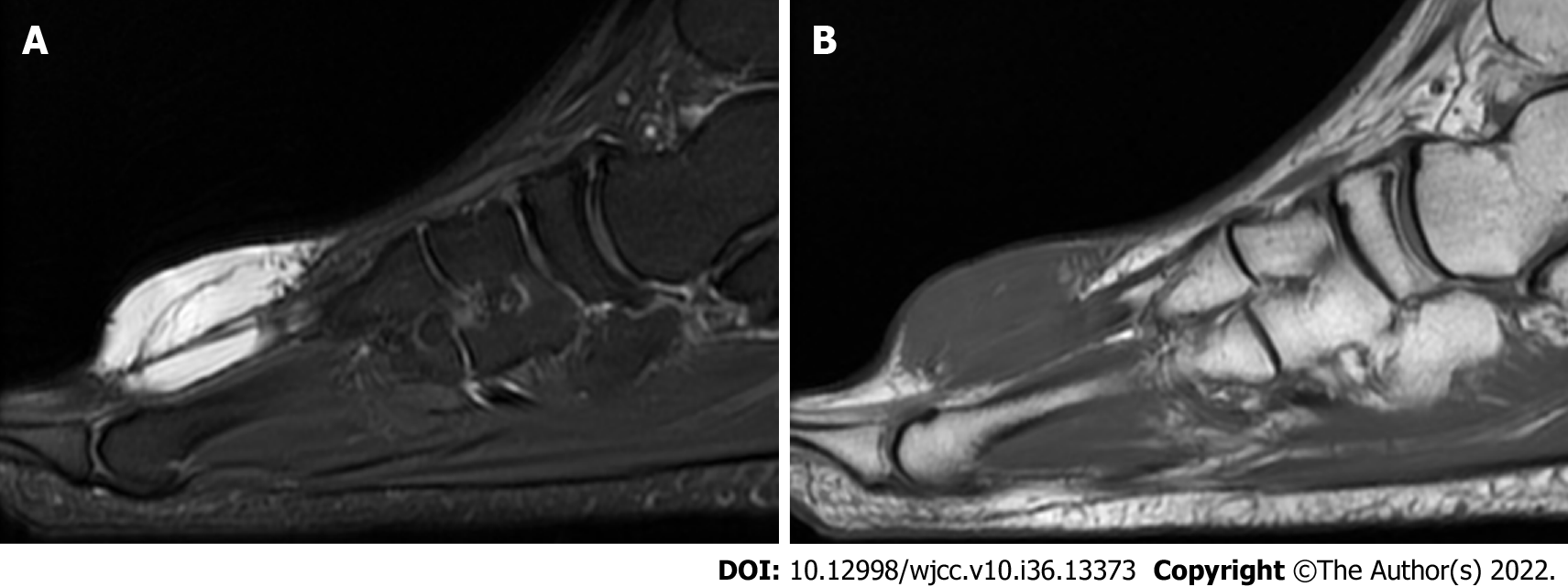
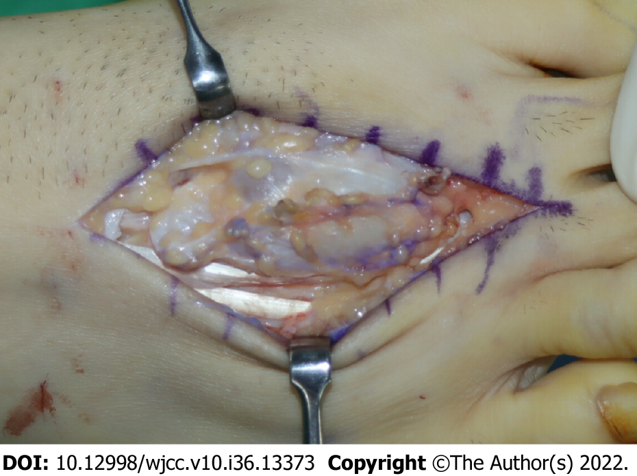
Figure 1 Multiple lobulated cystic lesions within and above the second extensor digitorum brevis tendon.
A: Fat-saturation T2-weighted image; B: Fat-saturation T1-weighted image.
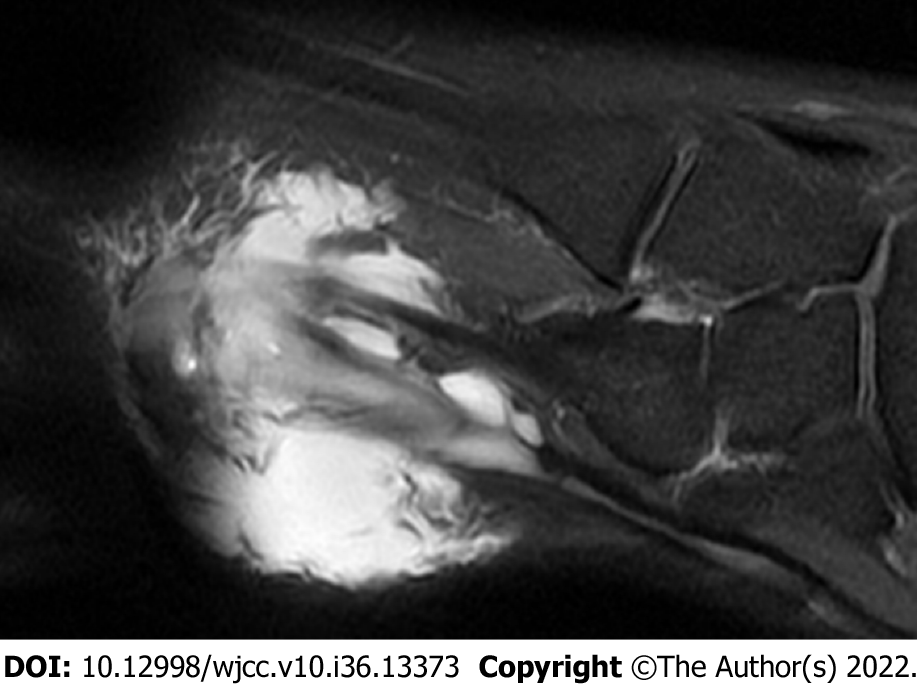
Figure 2 Extensive tenosynovitis around the ganglion cysts.
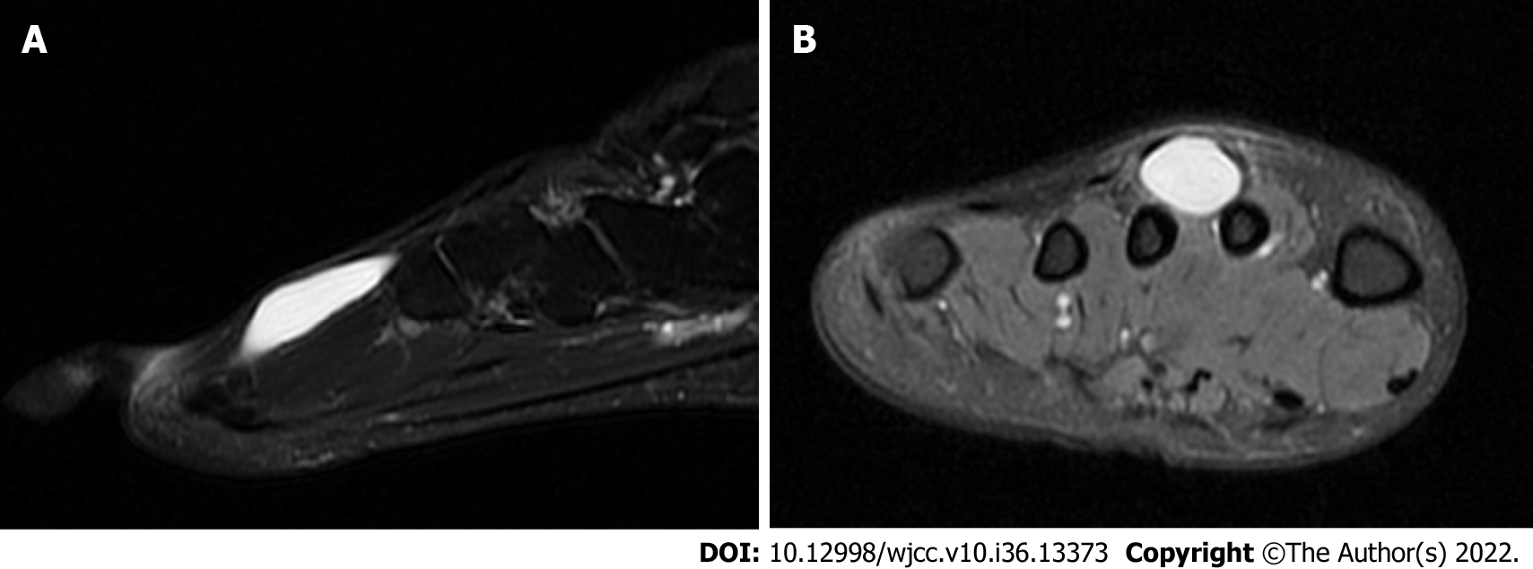
Figure 3 Intratendinous ganglion of the second extensor digitorum brevis tendon without extra-tendinous lesion and tenosynovitis on a fat-saturation T2-weighted image.
A: Sagittal image; B: Axial image.
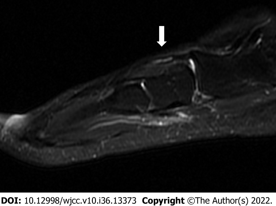
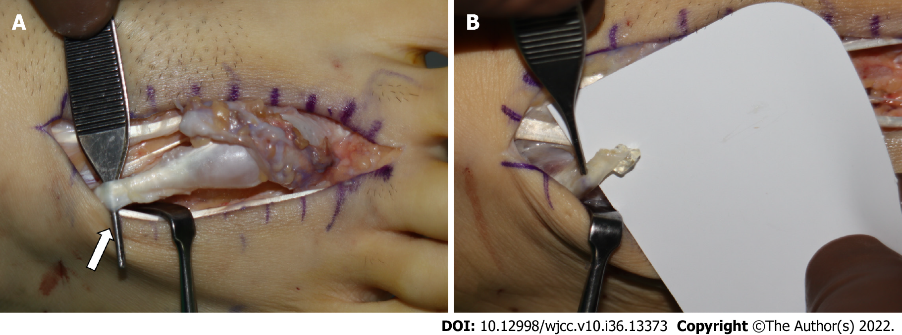
Figure 4 Satellite lesion at the proximal end of the second extensor digitorum brevis tendon before attaching to the calcaneus.
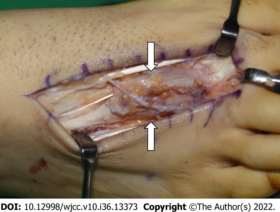
Figure 5 Extra-tendinous ganglion cysts and tenosynovitis over the extensor tendons.
Figure 6 The second extensor digitorum brevis tendon with enlarged spindle shape.
Figure 7 Satellite lesion of the second extensor digitorum brevis tendon.
A: Bulbous satellite lesions in the proximal portion of the tendon; B: Colorless jelly-like content from the inside of the incised tendon.
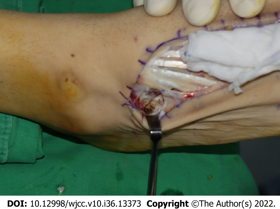
Figure 8 Remnant tendon removal and electrocauterization were performed at the anterior process of the calcaneus.
- Citation: Park JJ, Seok HG, Yan H, Park CH. Recurrence of intratendinous ganglion due to incomplete excision of satellite lesion in the extensor digitorum brevis tendon: A case report. World J Clin Cases 2022; 10(36): 13373-13380
- URL: https://www.wjgnet.com/2307-8960/full/v10/i36/13373.htm
- DOI: https://dx.doi.org/10.12998/wjcc.v10.i36.13373


