©The Author(s) 2022.
World J Clin Cases. Dec 6, 2022; 10(34): 12684-12689
Published online Dec 6, 2022. doi: 10.12998/wjcc.v10.i34.12684
Published online Dec 6, 2022. doi: 10.12998/wjcc.v10.i34.12684
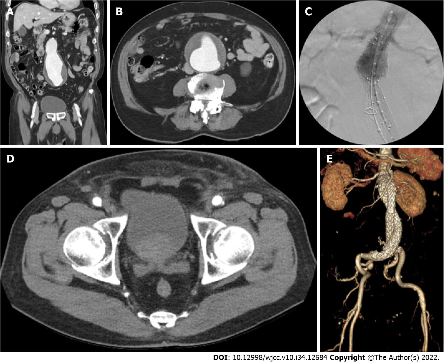
Figure 1 computed tomography scan of the patient with an abdominal aortic aneurysm.
A: Coronal view of the preoperative computed tomography (CT); B: Cross-sectional view of the preoperative CT; C: Intraoperative completion angiogram of the aortic neck; D: Cross-sectional view of the bilateral access sites of the postoperative CT scan: E: Volume-rendered image of the postoperative CT scan.
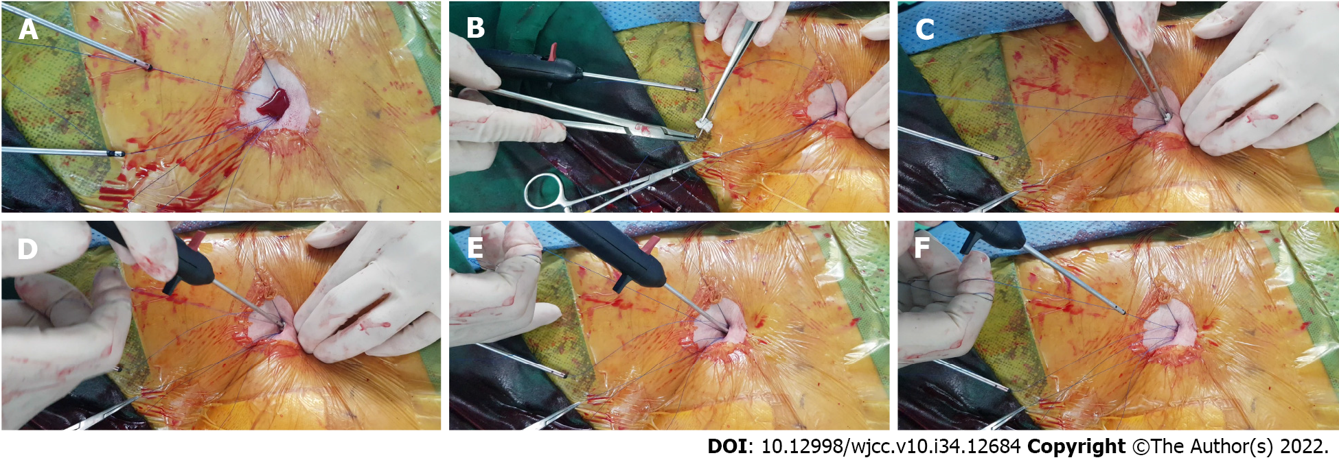
Figure 2 Surgical-assisted hemostasis technique.
A: After the successful deployment of the 2 ProGlide, substantial oozing was observed at the access site; B: Small piece of rolled Surgicel was sutured over the ProGlide suture thread through the empty needle; C: The attached Surgicel was placed at the access-site opening; D: The Surgicel was inserted onto the arterial wall along the suture thread with the pusher, E: Mild compression was applied with the pusher; F: Complete hemostasis was achieved.
- Citation: Kim H, Lee K, Cho S, Joh JH. Rapid hemostasis of the residual inguinal access sites during endovascular procedures: A case report. World J Clin Cases 2022; 10(34): 12684-12689
- URL: https://www.wjgnet.com/2307-8960/full/v10/i34/12684.htm
- DOI: https://dx.doi.org/10.12998/wjcc.v10.i34.12684


