Copyright
©The Author(s) 2022.
World J Clin Cases. Nov 26, 2022; 10(33): 12345-12351
Published online Nov 26, 2022. doi: 10.12998/wjcc.v10.i33.12345
Published online Nov 26, 2022. doi: 10.12998/wjcc.v10.i33.12345
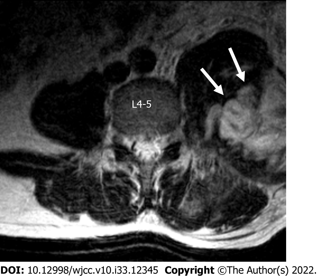
Figure 1 Axial T2-weighted magnetic resonance image scan of the lumbar spine showed left psoas muscle hematoma (arrows).
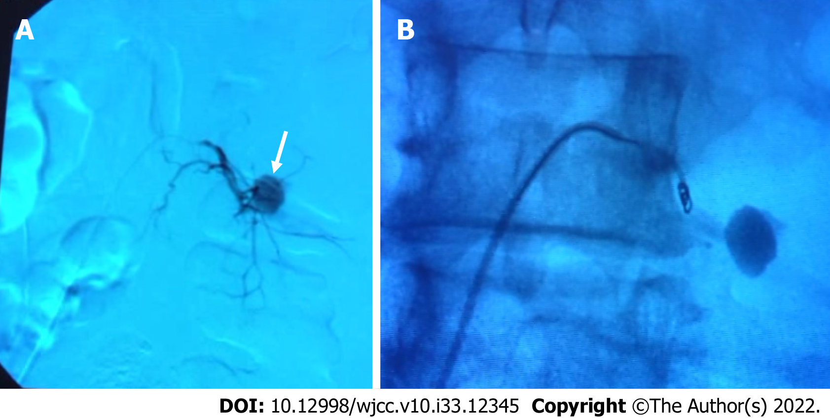
Figure 2 Arteriography.
A: Arteriography showed pseudoaneurysm (arrow) of the left 4th lumbar segmental artery; B: Arteriography of embolization using microcoils.
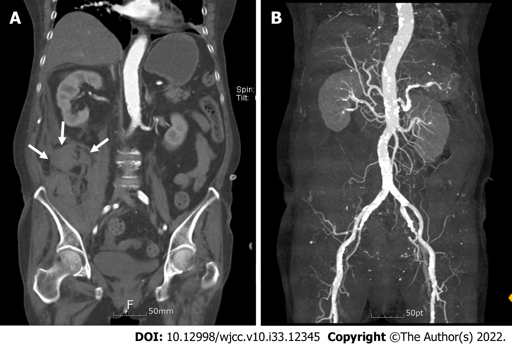
Figure 3 Computed tomography scans of the abdomen aorta.
A: Coronal image of the right retroperitoneal hemorrhage (arrows); B: Vascular reconstruction image showed no detected bleeding focus.
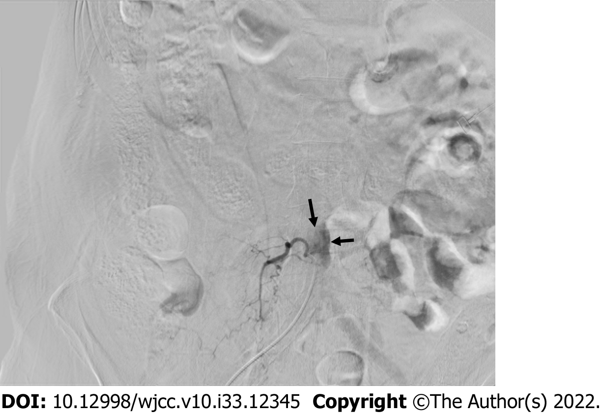
Figure 4 Arteriography showed the right 4th lumbar segmental artery rupture.
Arrows indicate blood leakage.
- Citation: Cho WJ, Kim KW, Park HY, Kim BH, Lee JS. Segmental artery injury during transforaminal percutaneous endoscopic lumbar discectomy: Two case reports. World J Clin Cases 2022; 10(33): 12345-12351
- URL: https://www.wjgnet.com/2307-8960/full/v10/i33/12345.htm
- DOI: https://dx.doi.org/10.12998/wjcc.v10.i33.12345


