Copyright
©The Author(s) 2022.
World J Clin Cases. Nov 16, 2022; 10(32): 12045-12051
Published online Nov 16, 2022. doi: 10.12998/wjcc.v10.i32.12045
Published online Nov 16, 2022. doi: 10.12998/wjcc.v10.i32.12045
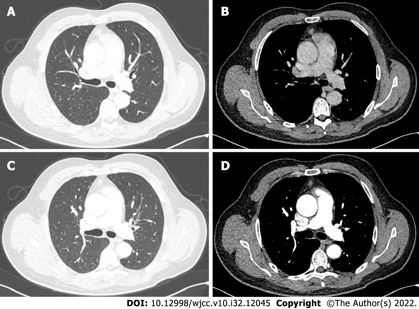
Figure 1 Imaging and macroscopic examination of the anterior mediastinum occupancy.
A and B: Plain computed tomography (CT) of pulmonary window (A) and mediastinal window (B) showed anterior mediastinal mass; C and D: The enhanced CT of pulmonary window (C) and mediastinal window (D) showed no significant enhancement of the anterior mediastinal mass.
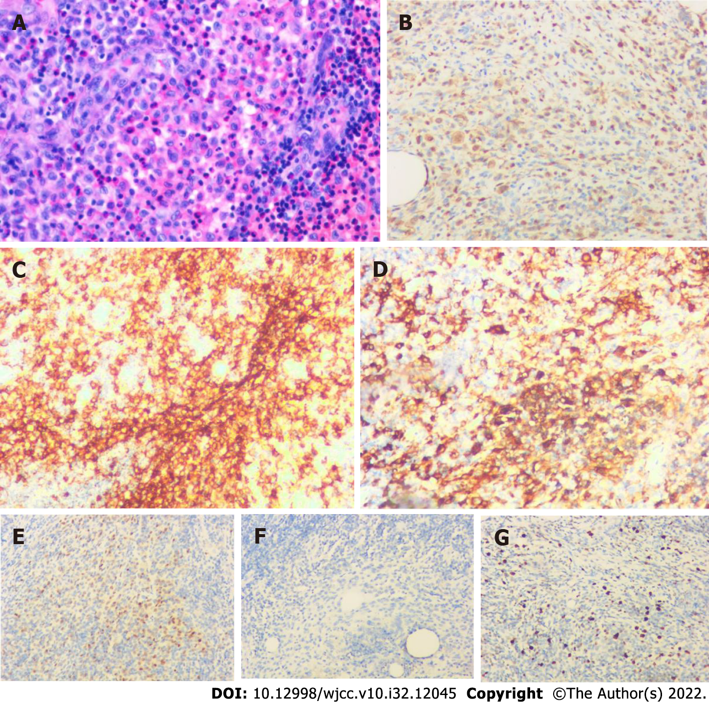
Figure 2 Histopathological analysis and immunohistochemical examination of the resected specimen.
A: Hematoxylin and eosin staining (200 ×); B-G: Immunohistochemical staining for (B) S100 (200 ×), (C) CD1a (200 ×), (D) CD163 (200 ×), (E) cyclin D1 (200 ×), (F) CD21 (200 ×) and (G) Ki-67 (200 ×).
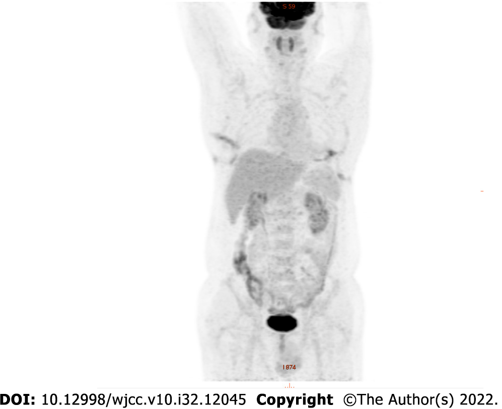
Figure 3 18F-fluorodeoxyglucose positron emission tomography showed positive uptake of the mass, suggesting no systemic metastasis.
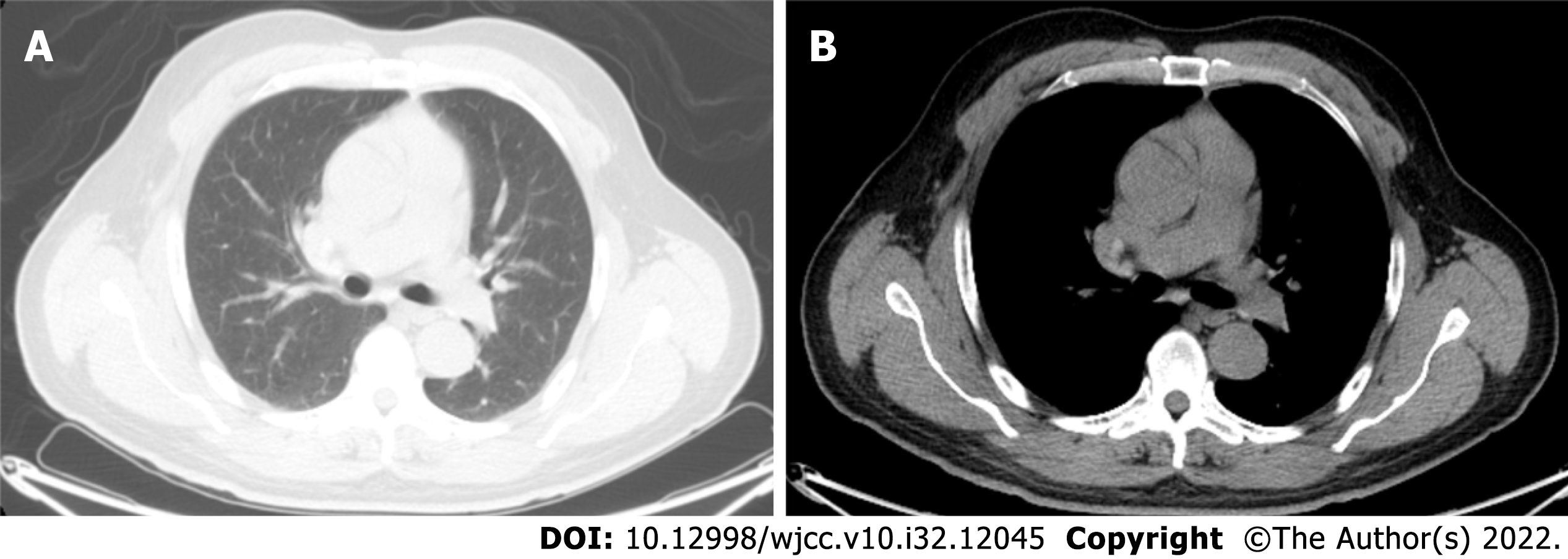
Figure 4 Computed tomography of the chest at 3 mo after surgery.
A: Plain Computed tomography (CT) of pulmonary window showed good postoperative healing; B: Plain CT of mediastinal window.
- Citation: Li YF, Han SH, Qie P, Yin QF, Wang HE. Langerhans cell histiocytosis involving only the thymus in an adult: A case report. World J Clin Cases 2022; 10(32): 12045-12051
- URL: https://www.wjgnet.com/2307-8960/full/v10/i32/12045.htm
- DOI: https://dx.doi.org/10.12998/wjcc.v10.i32.12045


