Copyright
©The Author(s) 2022.
World J Clin Cases. Nov 16, 2022; 10(32): 11942-11948
Published online Nov 16, 2022. doi: 10.12998/wjcc.v10.i32.11942
Published online Nov 16, 2022. doi: 10.12998/wjcc.v10.i32.11942
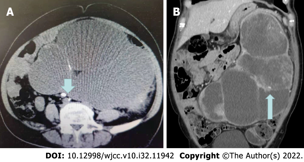
Figure 1 Computed tomography.
A: Computed tomography (CT) image showing a ureter stone (arrow); B: CT image showing renal parenchyma masses (arrow).
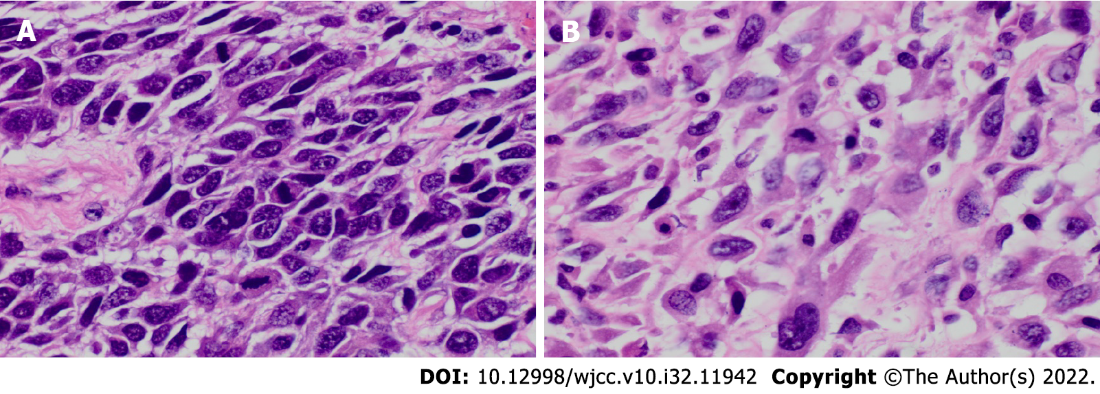
Figure 2 Pathological results of renal mass after resection.
A: Squamous cell carcinoma area (hematoxylin-eosin, 400 ×); B: Dedifferentiated sarcomatosis area (hematoxylin-eosin, 400 ×).
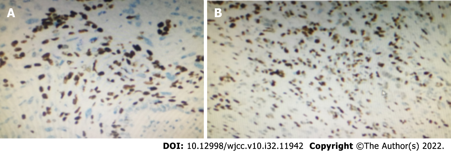
Figure 3 Immunohistochemistry revealed.
A: P40 (small focus squamous area +); B: P63 (squamous area and diffuse area +).
- Citation: Liu XH, Zou QM, Cao JD, Wang ZC. Primary squamous cell carcinoma with sarcomatoid differentiation of the kidney associated with ureteral stone obstruction: A case report . World J Clin Cases 2022; 10(32): 11942-11948
- URL: https://www.wjgnet.com/2307-8960/full/v10/i32/11942.htm
- DOI: https://dx.doi.org/10.12998/wjcc.v10.i32.11942


