Copyright
©The Author(s) 2022.
World J Clin Cases. Nov 16, 2022; 10(32): 11861-11868
Published online Nov 16, 2022. doi: 10.12998/wjcc.v10.i32.11861
Published online Nov 16, 2022. doi: 10.12998/wjcc.v10.i32.11861
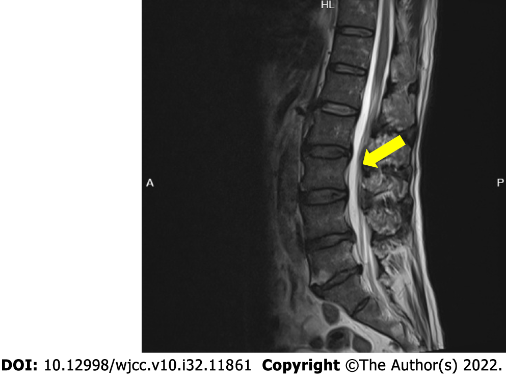
Figure 1 Magnetic resonance imaging showing disc herniations at L3/4, L4/5, and L5/S1 (arrow).
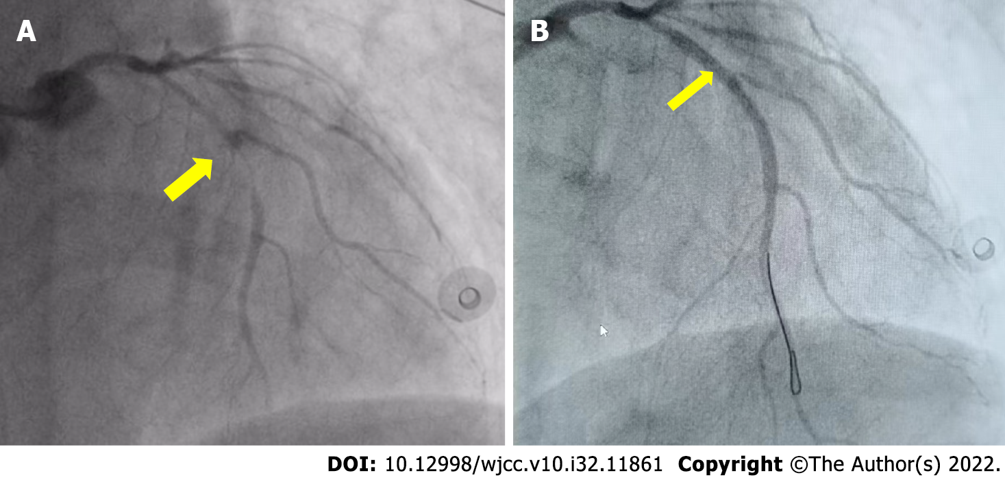
Figure 2 Percutaneous coronary angiography.
A: Before percutaneous coronary intervention; B: After percutaneous coronary intervention.
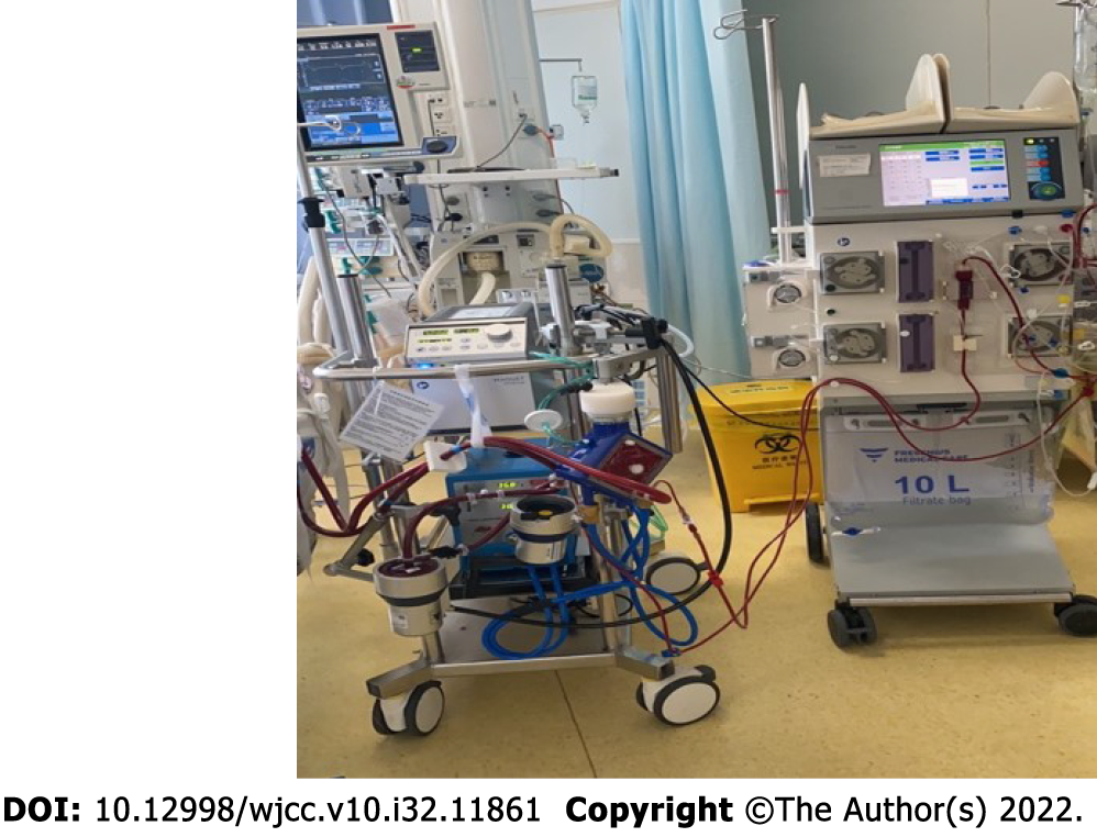
Figure 3 Equipment used for extracorporeal membrane oxygenation and continuous renal replacement therapy.
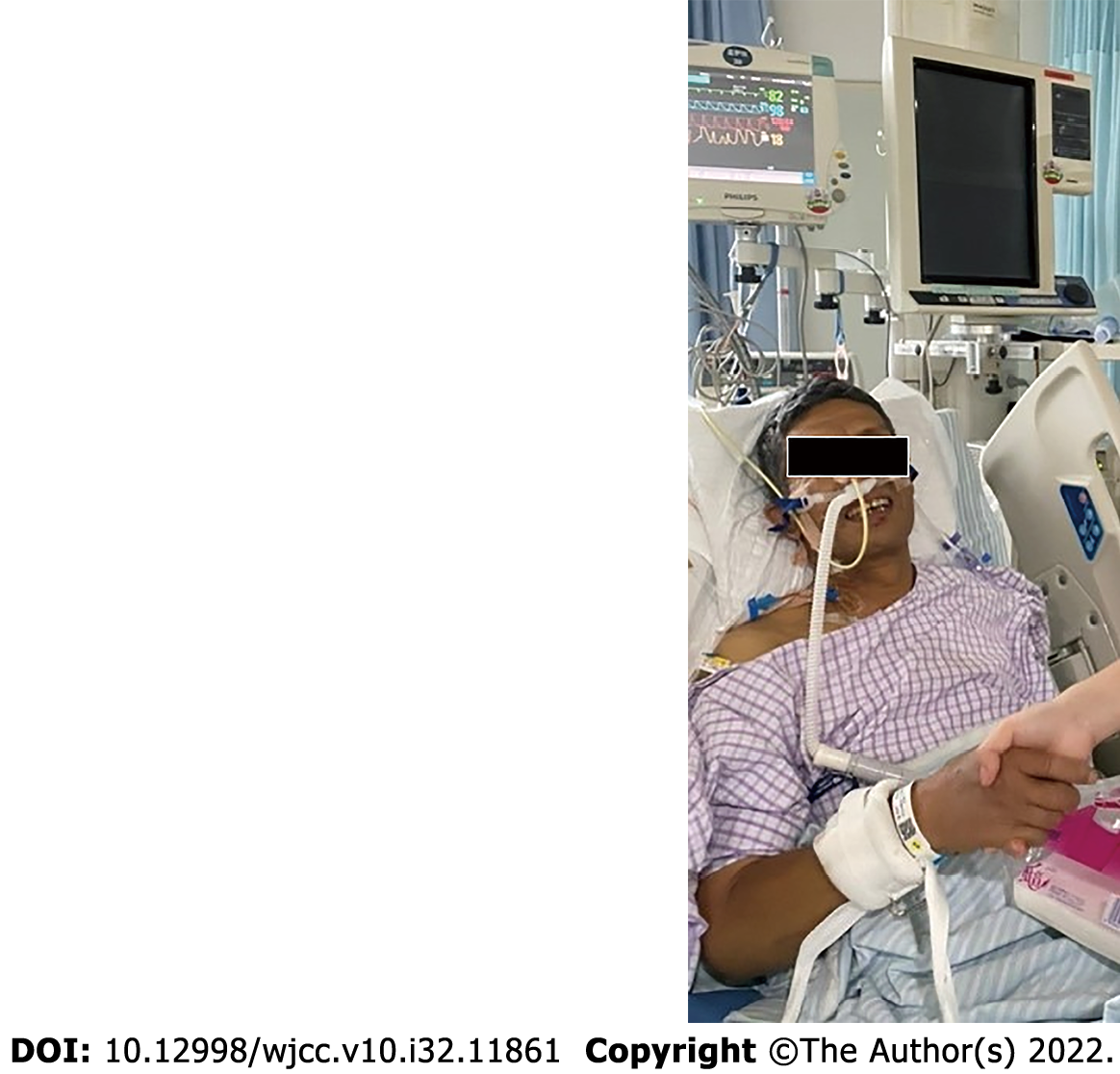
Figure 4 The patient after successful removal of tracheal intubation.
- Citation: Wang QQ, Jiang Y, Zhu JG, Zhang LW, Tong HJ, Shen P. Survival of a patient who received extracorporeal membrane oxygenation due to postoperative myocardial infarction: A case report. World J Clin Cases 2022; 10(32): 11861-11868
- URL: https://www.wjgnet.com/2307-8960/full/v10/i32/11861.htm
- DOI: https://dx.doi.org/10.12998/wjcc.v10.i32.11861


