Copyright
©The Author(s) 2022.
World J Clin Cases. Nov 6, 2022; 10(31): 11549-11554
Published online Nov 6, 2022. doi: 10.12998/wjcc.v10.i31.11549
Published online Nov 6, 2022. doi: 10.12998/wjcc.v10.i31.11549
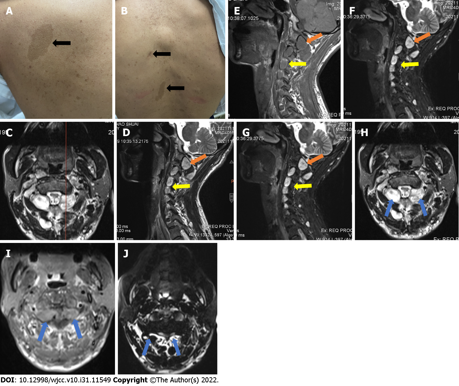
Figure 1 The physical signs and imaging data pre-operation.
A: Representative café-au-lait spots near the left scapula and on the back (B); C-J: Preoperative MRI images of the patient, showing conspicuous neoplasms in the upper cervical spine (yellow and blue arrows) and the presence of nodules at C2-7 (yellow arrows); C: At the position line of the sagittal and coronal planes, the neoplasm presents high signal on T1 (D, H), relatively low signal on T2 (E, I); signal enhancement showed high intensity in all nodules (F, J) images.
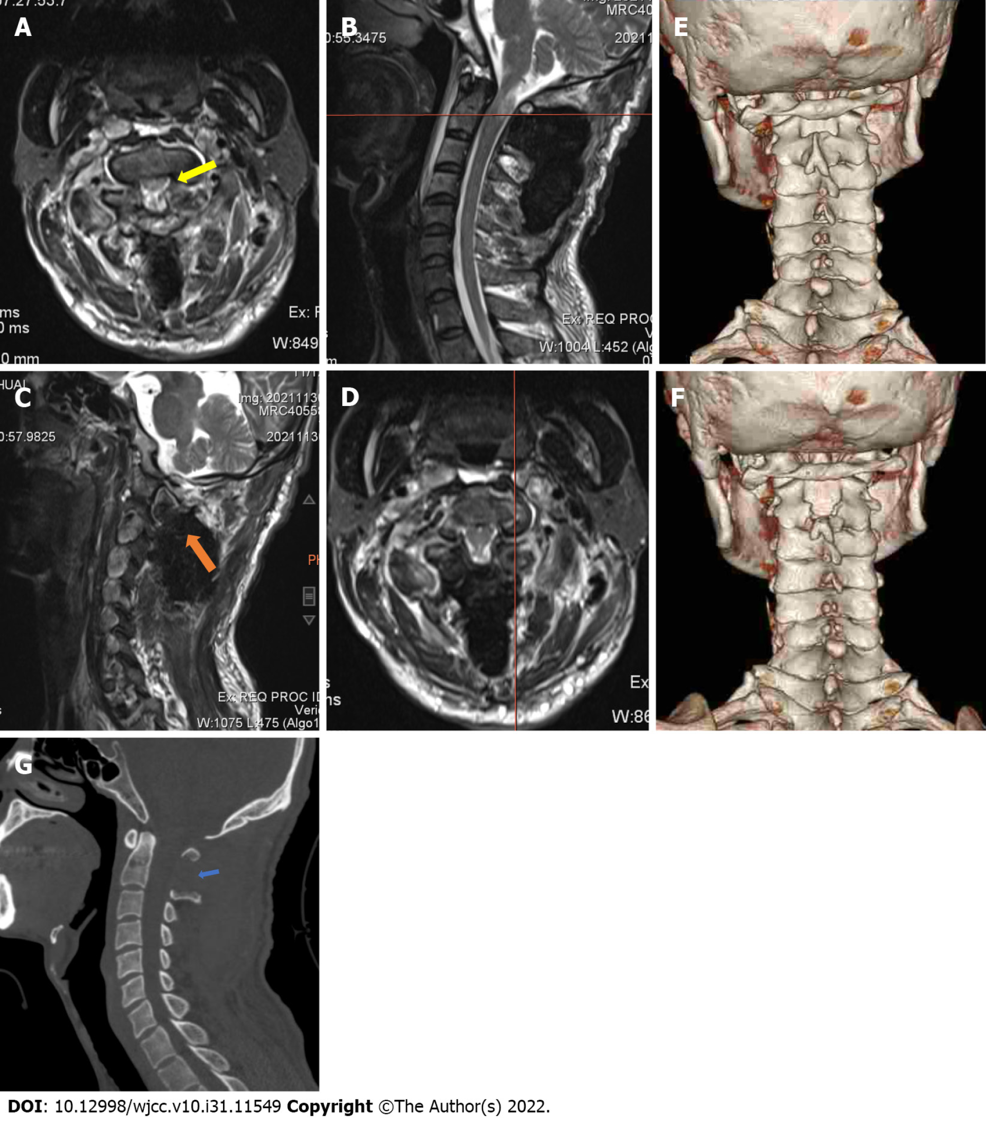
Figure 2 Post-operative images of the patient.
A: The coronal plane showed complete removal of the neoplasm and relief from compression of the spinal cord (yellow arrow) at the C1 position line (B); C: The sagittal plane also showed removal of the neoplasm (orange arrow) at the position line (D); Comparison of the pre-operative (E) and post-operative (F) images to three dimensional computed tomography showed limited structural changes on the posterior arch and laminar of the axis, with (G) substantial preservation of the upper cervical spine.
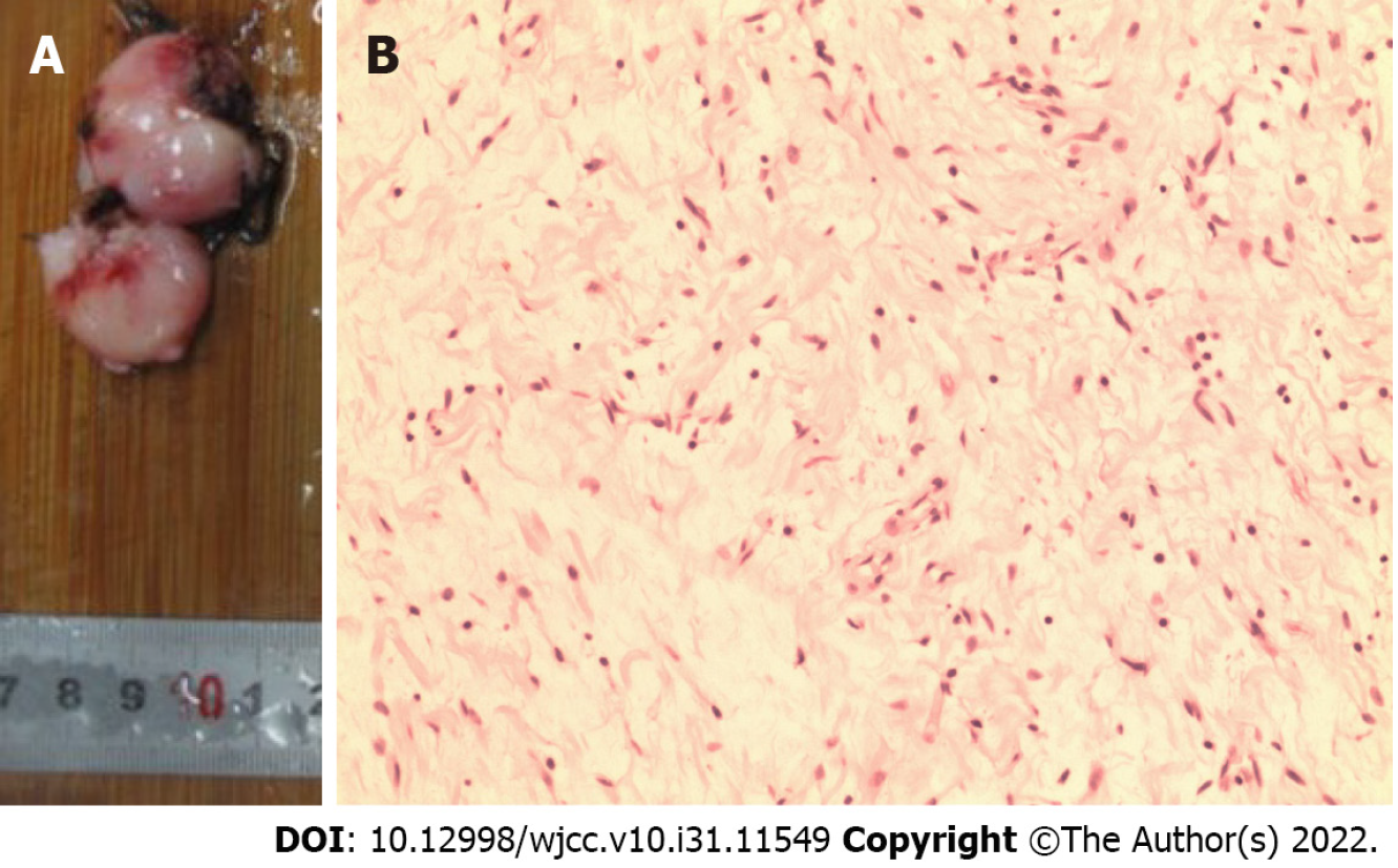
Figure 3 The picture of the resected tumor and the result of histopathological examination.
A: The tumor was resected intact as two parts; B: Histopathological examination revealed spindle ganglion cells spreading throughout the tumor, without pleomorphism.
- Citation: Wang S, Ma JX, Zheng L, Sun ST, Xiang LB, Chen Y. Multiple bilateral and symmetric C1-2 ganglioneuromas: A case report. World J Clin Cases 2022; 10(31): 11549-11554
- URL: https://www.wjgnet.com/2307-8960/full/v10/i31/11549.htm
- DOI: https://dx.doi.org/10.12998/wjcc.v10.i31.11549


