
















©The Author(s) 2022.
World J Clin Cases. Oct 16, 2022; 10(29): 10794-10802
Published online Oct 16, 2022. doi: 10.12998/wjcc.v10.i29.10794
Published online Oct 16, 2022. doi: 10.12998/wjcc.v10.i29.10794
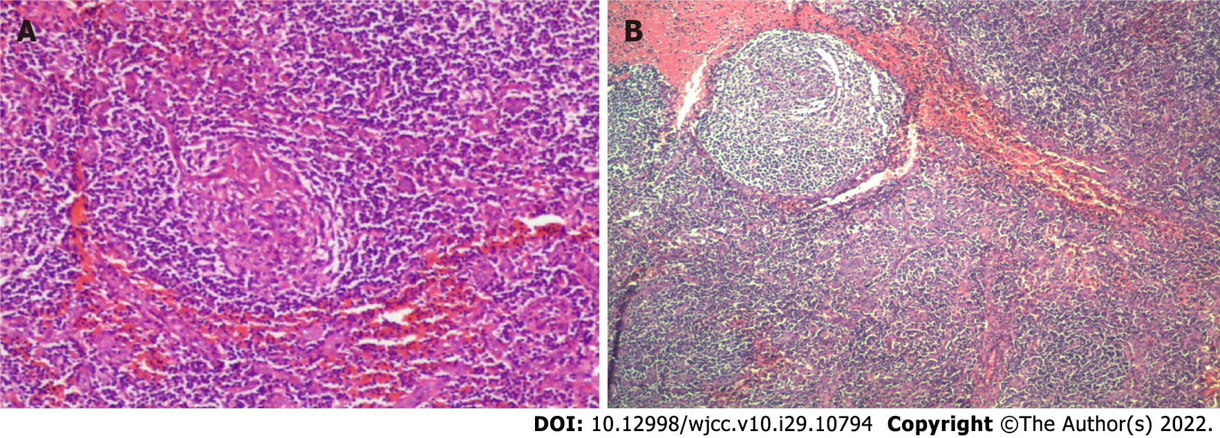
Figure 1 Benign hyperplasia of right axillary lymph nodes showed Castleman disease like changes.
A: Small lymph nodes submitted for examination showed a thin capsule, edema, marginal sinus, and lymphoid follicular-like structure in the node, obvious cell transformation in the germinal center, obvious vascular proliferation, mild proliferation of cells in the mantle area, partial "onion skin"-like structure, obvious proliferation in the T area (mainly proliferation of small lymphoid cells), an obvious increase in blood vessels, endothelial swelling and partial vitreous degeneration. Immunohistochemistry staining results: Ki-67 [granulosa cell (GC) +, the remaining 5% +], B cell lymphoma (GC -), Castleman's disease (CD) 3 (T region +), CD20 (focal +), CD21 (follicle dendritic cell +), CD10 (granulocyte +, GC weak +), CD138 (small amount +), human herpesvirus-8 (-); In situ hybridization results: Epstein-Barr virus in-situ hybridization (-). (Hematoxylin-eosin staining × 10); B: The right axillary lymph nodes showed reactive hyperplasia without tumor and tuberculosis. As observed with a light mirror.
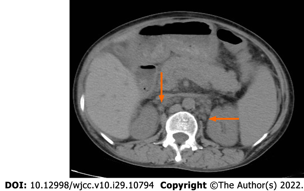
Figure 2 Abdominal computed tomography.
The number of lymph nodes in the abdominal cavity, retroperitoneum, and bilateral inguinal areas increased. The orange arrow shows the enlarged lymph nodes in the abdominal cavity.
- Citation: Liu XR, Tian M. Glucocorticoids combined with tofacitinib in the treatment of Castleman's disease: A case report. World J Clin Cases 2022; 10(29): 10794-10802
- URL: https://www.wjgnet.com/2307-8960/full/v10/i29/10794.htm
- DOI: https://dx.doi.org/10.12998/wjcc.v10.i29.10794