Copyright
©The Author(s) 2022.
World J Clin Cases. Oct 16, 2022; 10(29): 10695-10700
Published online Oct 16, 2022. doi: 10.12998/wjcc.v10.i29.10695
Published online Oct 16, 2022. doi: 10.12998/wjcc.v10.i29.10695
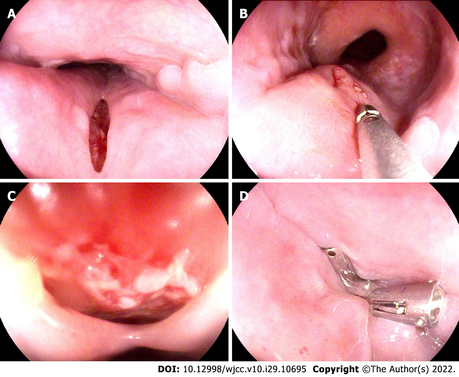
Figure 1 Resolution of the laceration above the mass with metal endoscopic clips.
A: 2 cm laceration of the esophagus (30 cm distal to the incisors); B: Submucosal mass was beneath the laceration, with spontaneous rupture; C: Detailed view of the crevasse showing granulated tissues and purulent exudate; D: Laceration was completely closed with metal endoscopic clips.
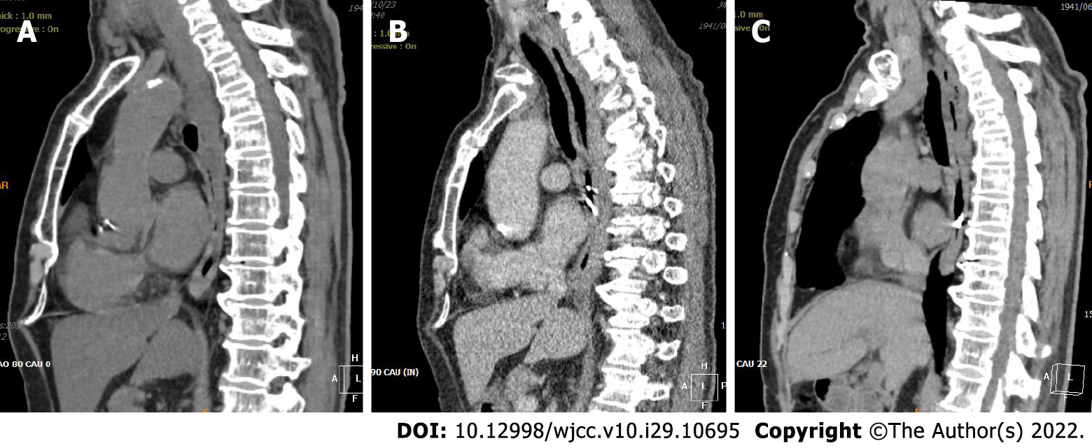
Figure 2 Computed tomography showed a double-barreled esophagus without thickening of the esophageal wall.
A: Chest computed tomography scan showed eccentric thickening of the esophageal wall; B: Chest computed tomography scan taken immediately after endoscopy showed worsened diffuse thickening of the esophageal wall; C: Chest computed tomography scan taken 2 mo after endoscopy showed that the thickening of the esophageal wall was alleviated with a double-barreled esophagus visible.
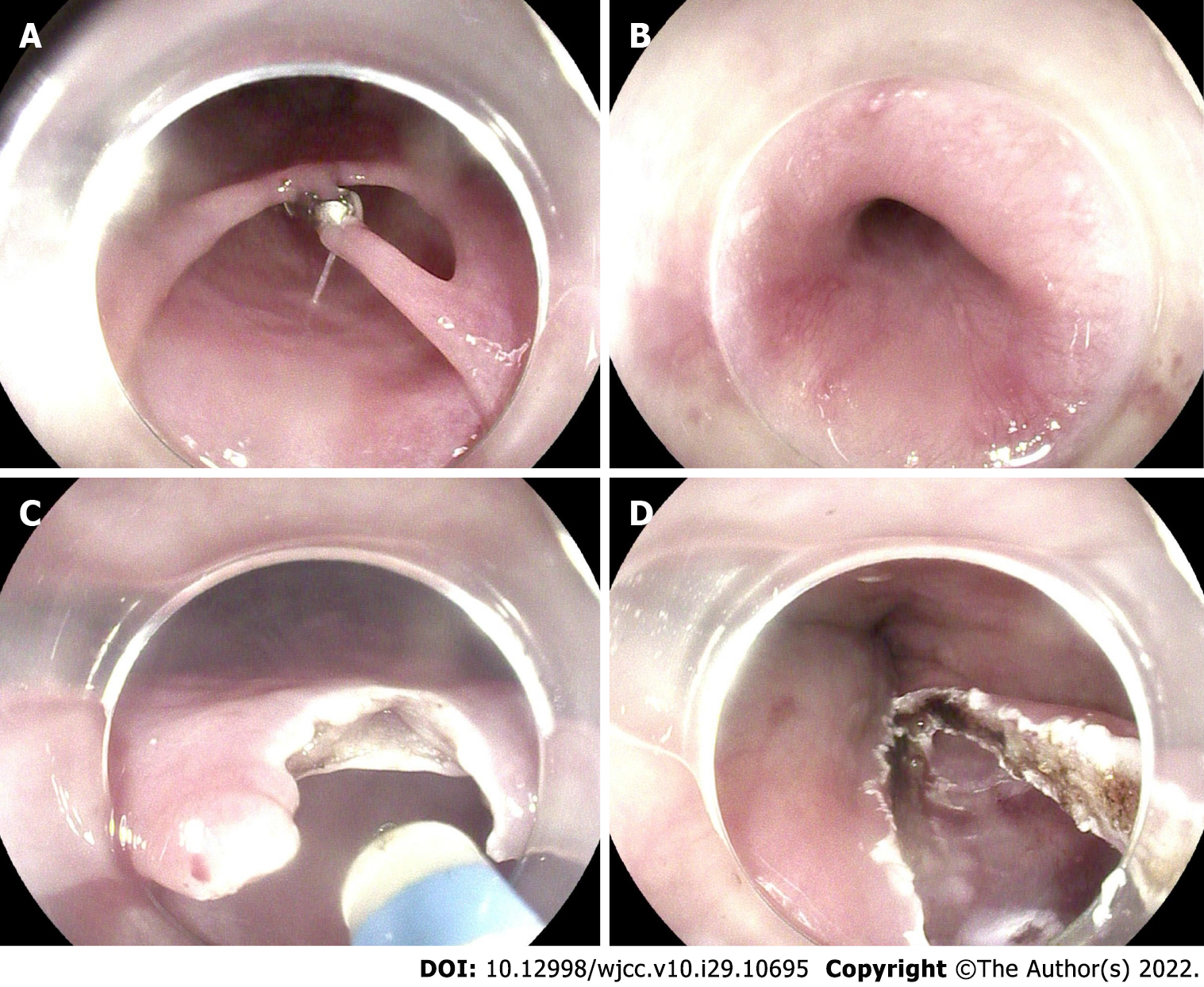
Figure 3 Endoscopic incision of the septum between two lumens was performed using a dual-knife process with diathermy.
A: Intramural submucosal dissection, with one endoscopic clip remaining; B: Internal space of the dissection; C: Endoscopic incision of the septum between two lumens; D: Completely cut septum.
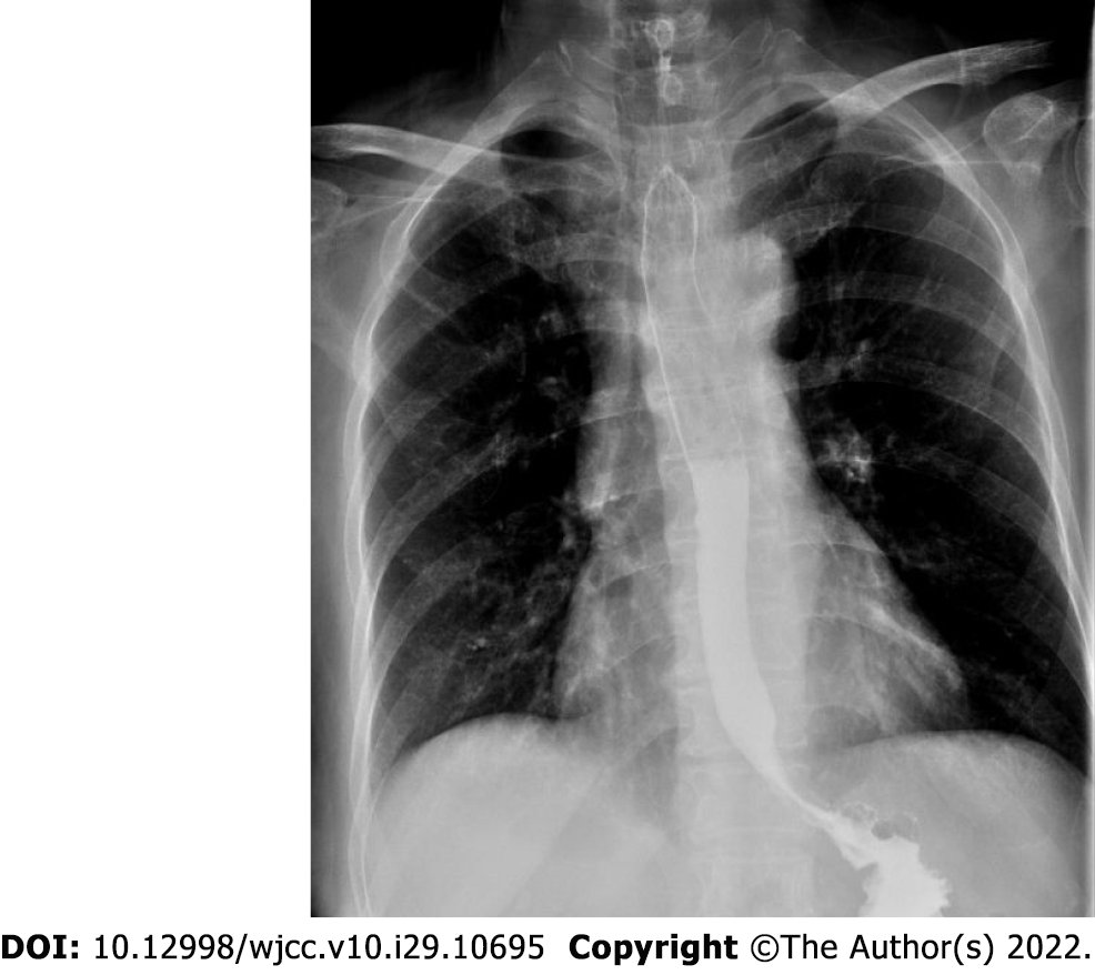
Figure 4 Esophagogram taken 3 d after endoscopic incision showed the dissection had disappeared, and the barium passed smoothly through the esophagus.
- Citation: Jiao Y, Sikong YH, Zhang AJ, Zuo XL, Gao PY, Ren QG, Li RY. Submucosal esophageal abscess evolving into intramural submucosal dissection: A case report. World J Clin Cases 2022; 10(29): 10695-10700
- URL: https://www.wjgnet.com/2307-8960/full/v10/i29/10695.htm
- DOI: https://dx.doi.org/10.12998/wjcc.v10.i29.10695


