Copyright
©The Author(s) 2022.
World J Clin Cases. Oct 6, 2022; 10(28): 10172-10179
Published online Oct 6, 2022. doi: 10.12998/wjcc.v10.i28.10172
Published online Oct 6, 2022. doi: 10.12998/wjcc.v10.i28.10172
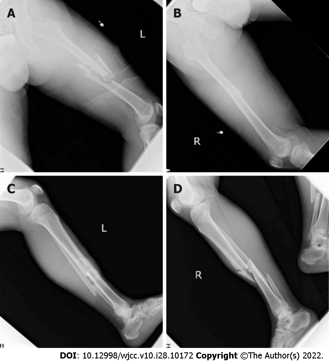
Figure 1 Initial plain radiographs revealed displaced bilateral femoral, tibial, and fibular midshaft fractures.
A and B: Bilateral femoral fractures; C and D: Tibial fracture.
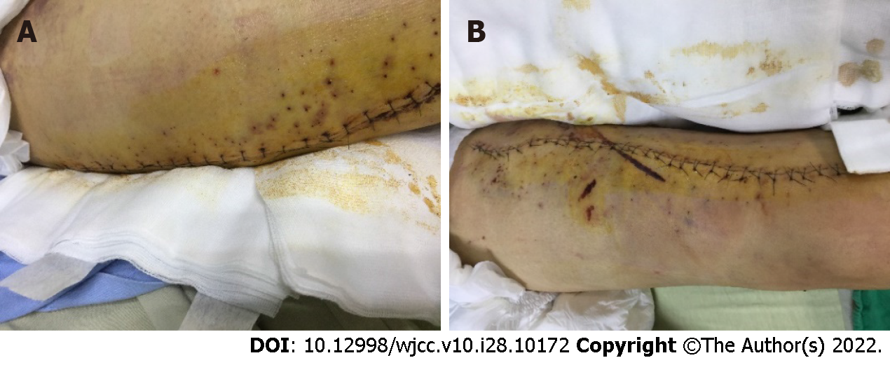
Figure 2 The photographs showed that we punctured hundreds of small holes around the closed wound using an 18-gauge needle in the thigh and leg, imitating a Chinese medicine bloodletting method, to allow the accumulated blood in the tissue to flow out to prevent skin necrosis and compartment syndrome.
A: Thigh; B: Leg.
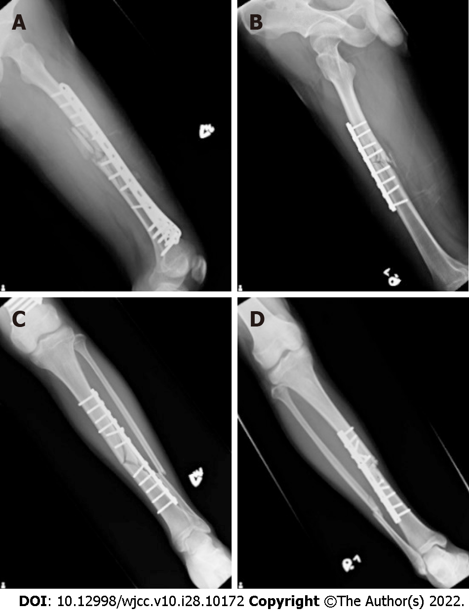
Figure 3 Postoperative X-rays illustrated that the patient received open reduction and internal fixation with one locking plate in the left femoral shaft, one broad dynamic compression plate in right femoral shaft, and two narrow dynamic compression plates in bilateral tibial shafts.
A: Left femoral shaft; B: Right femoral shaft; C and D: Two narrow dynamic compression plates in bilateral tibial shafts.
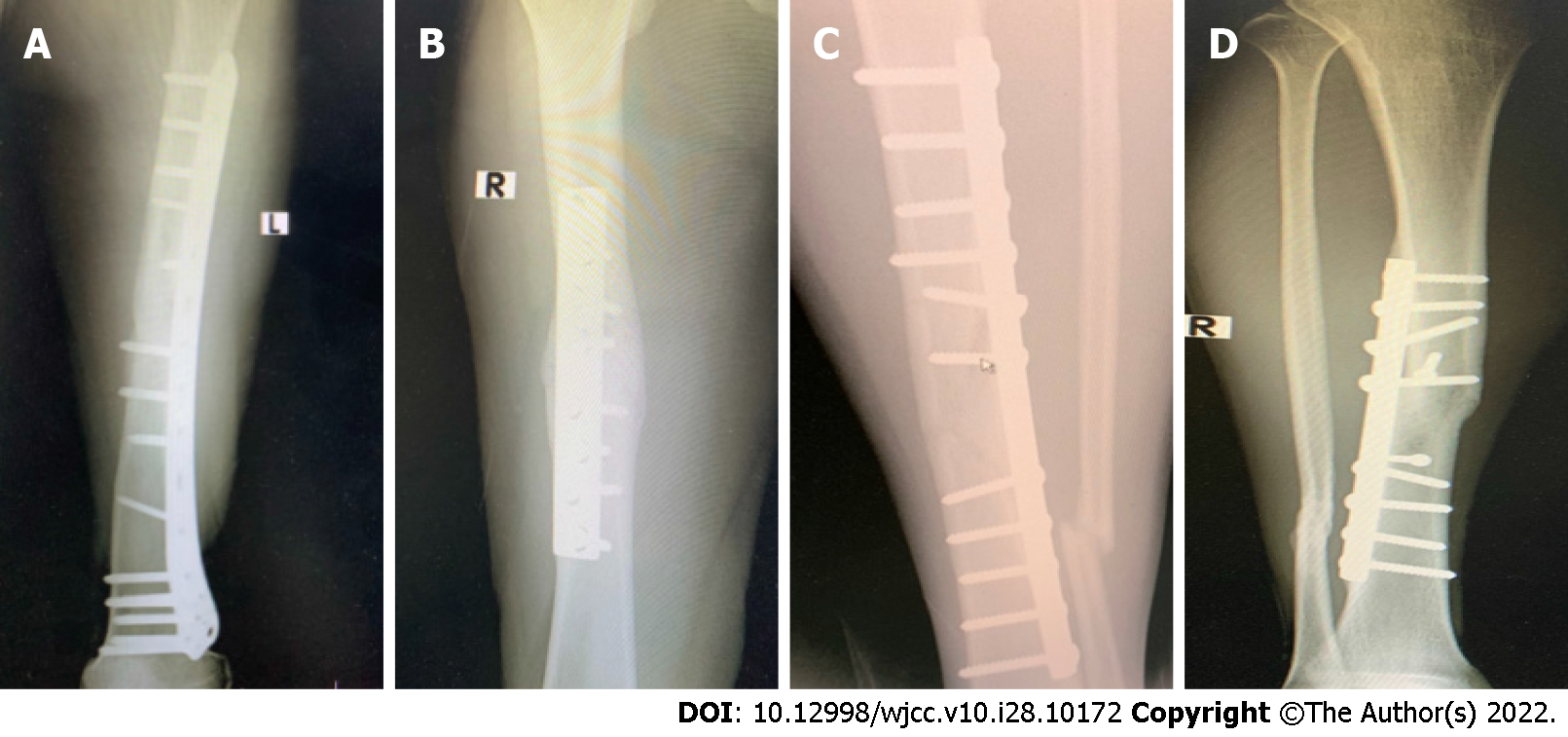
Figure 4 Radiography revealed bone union of bilateral femoral and tibial fracture sites at postoperative 13 mo.
A and B: Bone union of bilateral femoral; C and D: Bone union of tibial fracture.
- Citation: Wu CM, Liao HE, Lan SJ. Simultaneous bilateral floating knee: A case report. World J Clin Cases 2022; 10(28): 10172-10179
- URL: https://www.wjgnet.com/2307-8960/full/v10/i28/10172.htm
- DOI: https://dx.doi.org/10.12998/wjcc.v10.i28.10172


