Copyright
©The Author(s) 2022.
World J Clin Cases. Oct 6, 2022; 10(28): 10136-10145
Published online Oct 6, 2022. doi: 10.12998/wjcc.v10.i28.10136
Published online Oct 6, 2022. doi: 10.12998/wjcc.v10.i28.10136
Figure 1 Computed tomography scan.
A: Lesions in the liver, spleen, subcutaneous tissue (arrows); B: Lesions in the lungs (arrow); C: Pathological lymphadenopathy of the neck (arrow).
Figure 2 Upper endoscopy.
A: Atypical ulcers of the duodenum; B: Atypical polyps in gastric fundus (arrow); C: Atypical polyp in descending part of the duodenum.
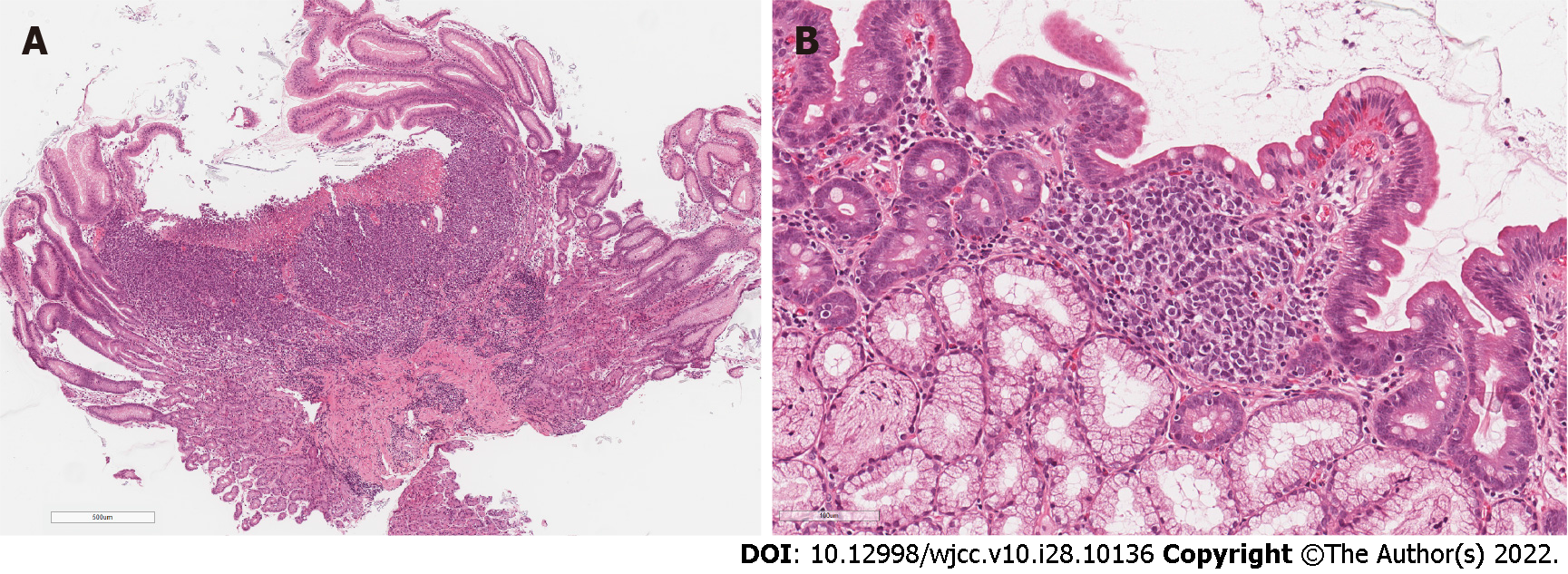
Figure 3 Stomach lesion and duodenal biopsy.
A: Stomach lesion biopsy. Ulcerated diffuse infiltrate in body-type mucosa; B: Duodenal biopsy. Subtle focal infiltrate in lamina propria, composed of medium-sized dyscohesive cells.
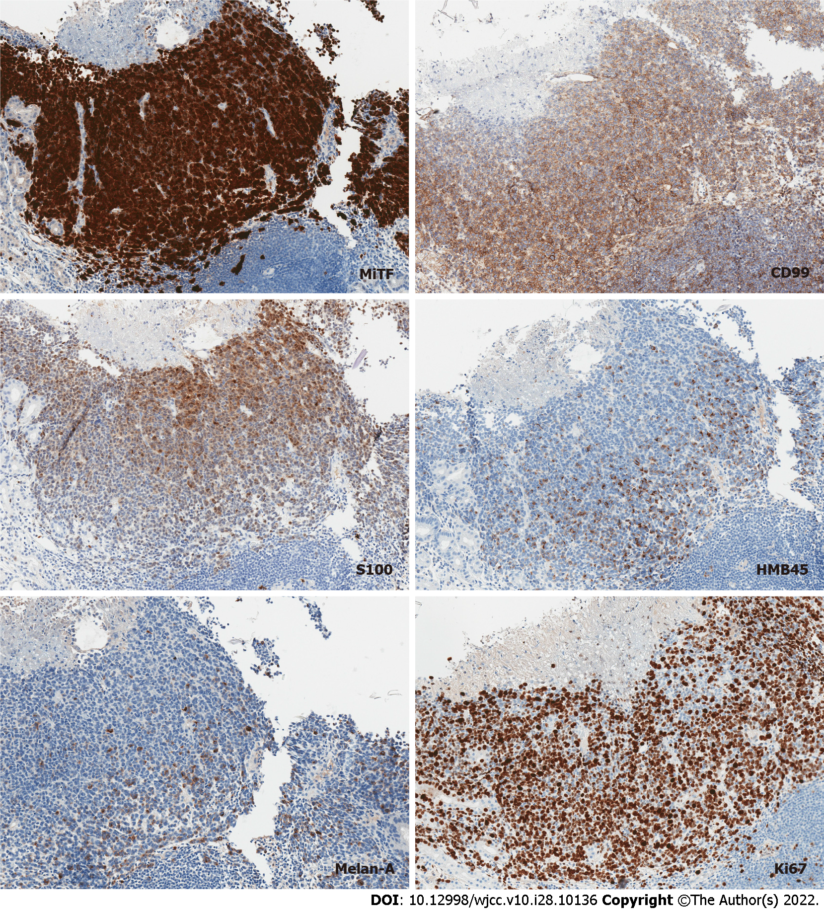
Figure 4 Immunohistochemistry.
Tumor cells diffusely and intensely positive for microphthalmia-associated transcription factor, diffusely positive for CD99, partially positive for S100 and focally faintly positive for HMB45 and Melan-A. Ki-67 proliferation index was up to 80%. MiTF: Microphthalmia-associated transcription factor.
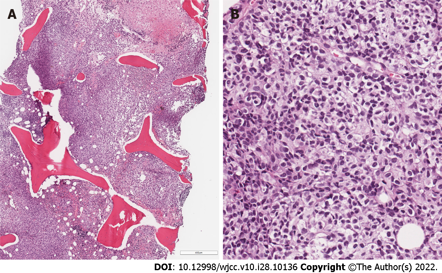
Figure 5 Bone marrow trephine biopsy.
A: Metastatic melanoma with focal necrosis replacing hematopoietic tissue; B: Diffuse compact infiltrate of epithelioid cells with clear or pale cytoplasm and oval, indented or elongated nuclei.
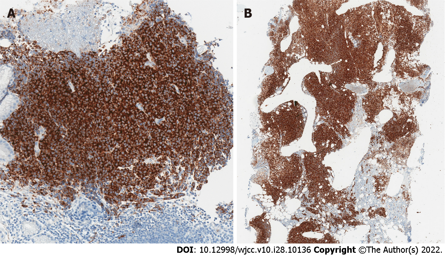
Figure 6 Strong diffuse tumor positivity for BRAF.
A: Gastric biopsy, B: Bone marrow trephine biopsy.
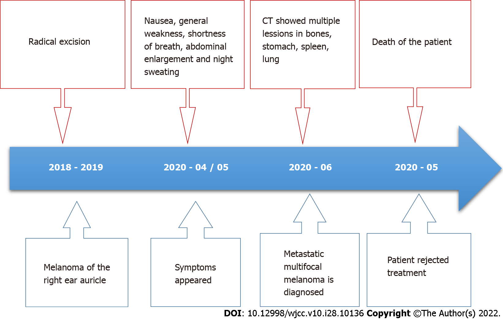
Figure 7 Case history timeline.
CT: Computed tomography.
- Citation: Maksimaityte V, Reivytyte R, Milaknyte G, Mickys U, Razanskiene G, Stundys D, Kazenaite E, Valantinas J, Stundiene I. Metastatic multifocal melanoma of multiple organ systems: A case report. World J Clin Cases 2022; 10(28): 10136-10145
- URL: https://www.wjgnet.com/2307-8960/full/v10/i28/10136.htm
- DOI: https://dx.doi.org/10.12998/wjcc.v10.i28.10136


