Copyright
©The Author(s) 2022.
World J Clin Cases. Sep 26, 2022; 10(27): 9790-9797
Published online Sep 26, 2022. doi: 10.12998/wjcc.v10.i27.9790
Published online Sep 26, 2022. doi: 10.12998/wjcc.v10.i27.9790
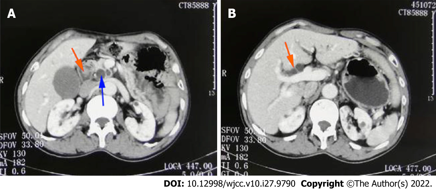
Figure 1 Contrast-enhanced computed tomography of the abdomen.
A: Space-occupying lesion in the duodenal papilla (orange arrowheads) and dilatation of the common bile duct (blue arrowheads); B: dilatation of extrahepatic bile duct (orange arrowheads).
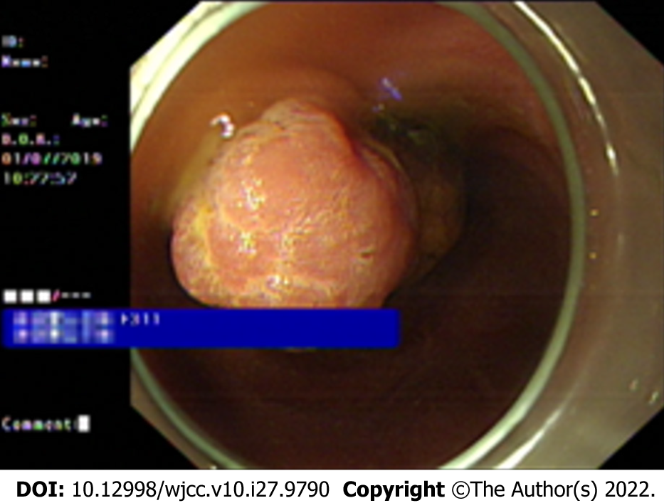
Figure 2 Endoscopic biopsy.
Tumor protruding from the duodenal papilla.
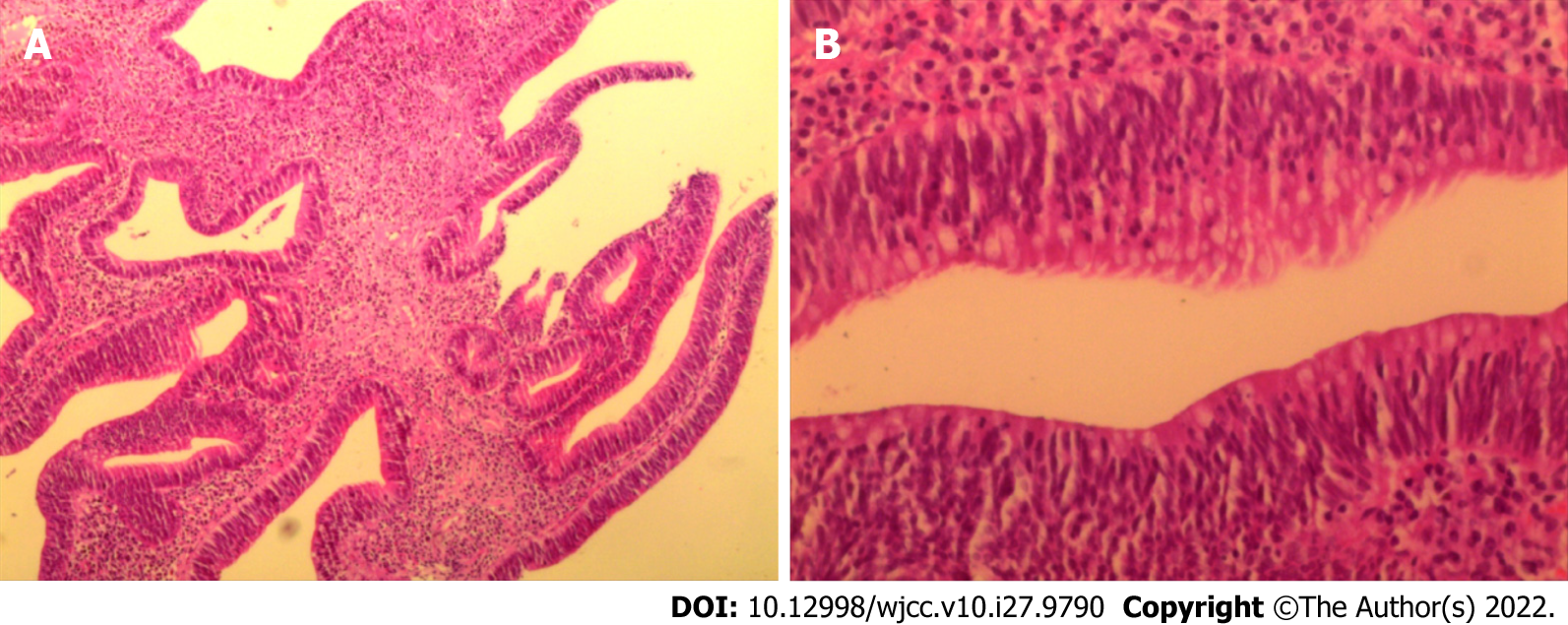
Figure 3 Histopathological findings of endoscopic biopsy.
A: Tubular villous growth. B: Moderate heterogeneous hyperplasia.
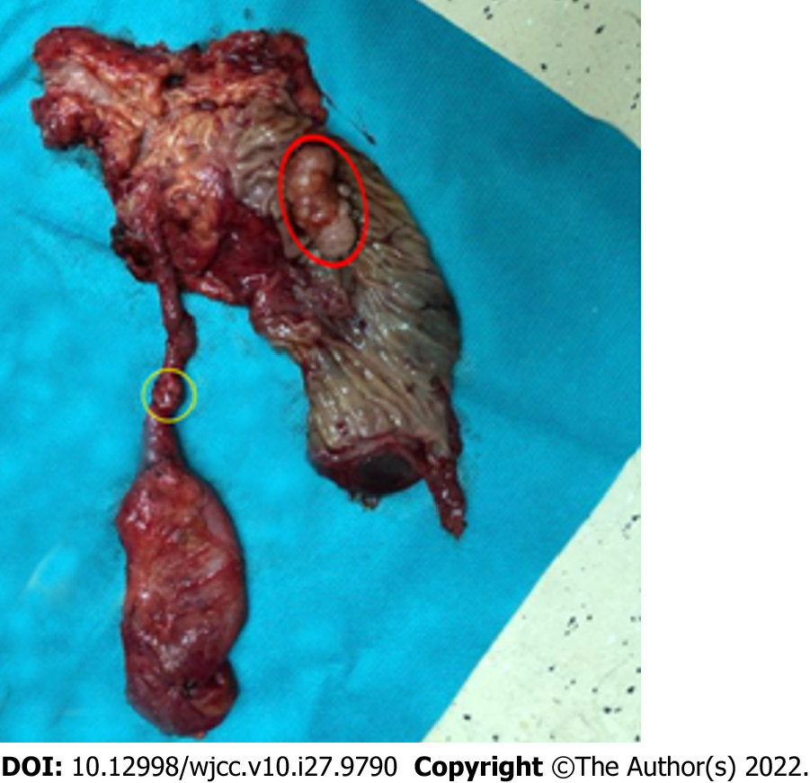
Figure 4 Macroscopic appearance of the surgical specimen.
Papillary tumor (red circle) and gallbladder neck duct tumor (yellow circle) were present in the specimen.
- Citation: Chen J, Zhu MY, Huang YH, Zhou ZC, Shen YY, Zhou Q, Fei MJ, Kong FC. Synchronous primary duodenal papillary adenocarcinoma and gallbladder carcinoma: A case report and review of literature. World J Clin Cases 2022; 10(27): 9790-9797
- URL: https://www.wjgnet.com/2307-8960/full/v10/i27/9790.htm
- DOI: https://dx.doi.org/10.12998/wjcc.v10.i27.9790


