Copyright
©The Author(s) 2022.
World J Clin Cases. Sep 26, 2022; 10(27): 9693-9702
Published online Sep 26, 2022. doi: 10.12998/wjcc.v10.i27.9693
Published online Sep 26, 2022. doi: 10.12998/wjcc.v10.i27.9693
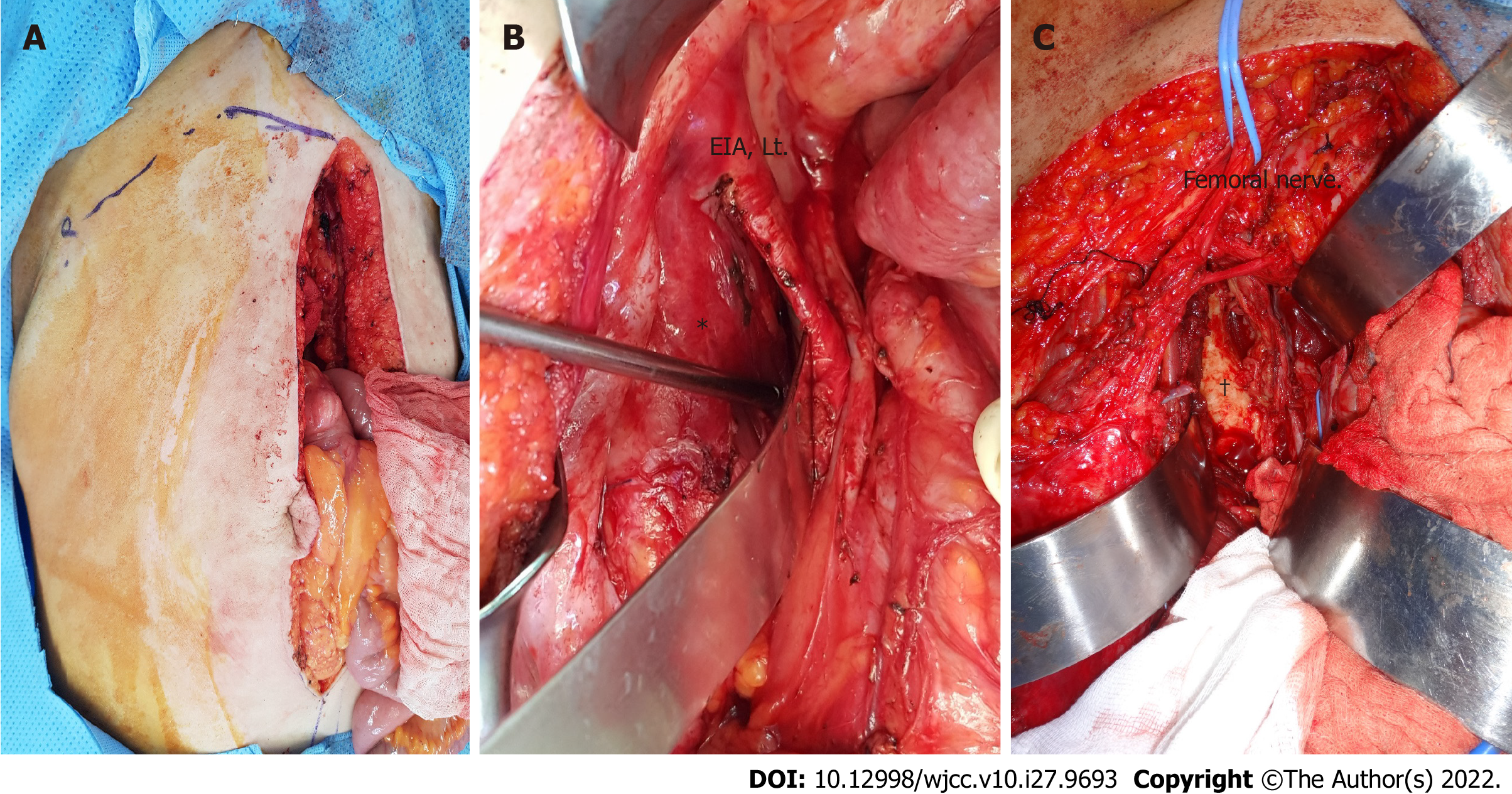
Figure 1 Intra and extra pelvic multidisciplinary surgical approach.
A: Incision of intra and extra pelvic approach (midline incision + ilioinguinal approach); B: Intra pelvic approach; C: Extrapelvic approach. *: Medial part of the sarcoma mass, soft tissue of the obturator internus; †: Lateral part of the sarcoma mass, from the ilium and ischium; EIA: External iliac artery.
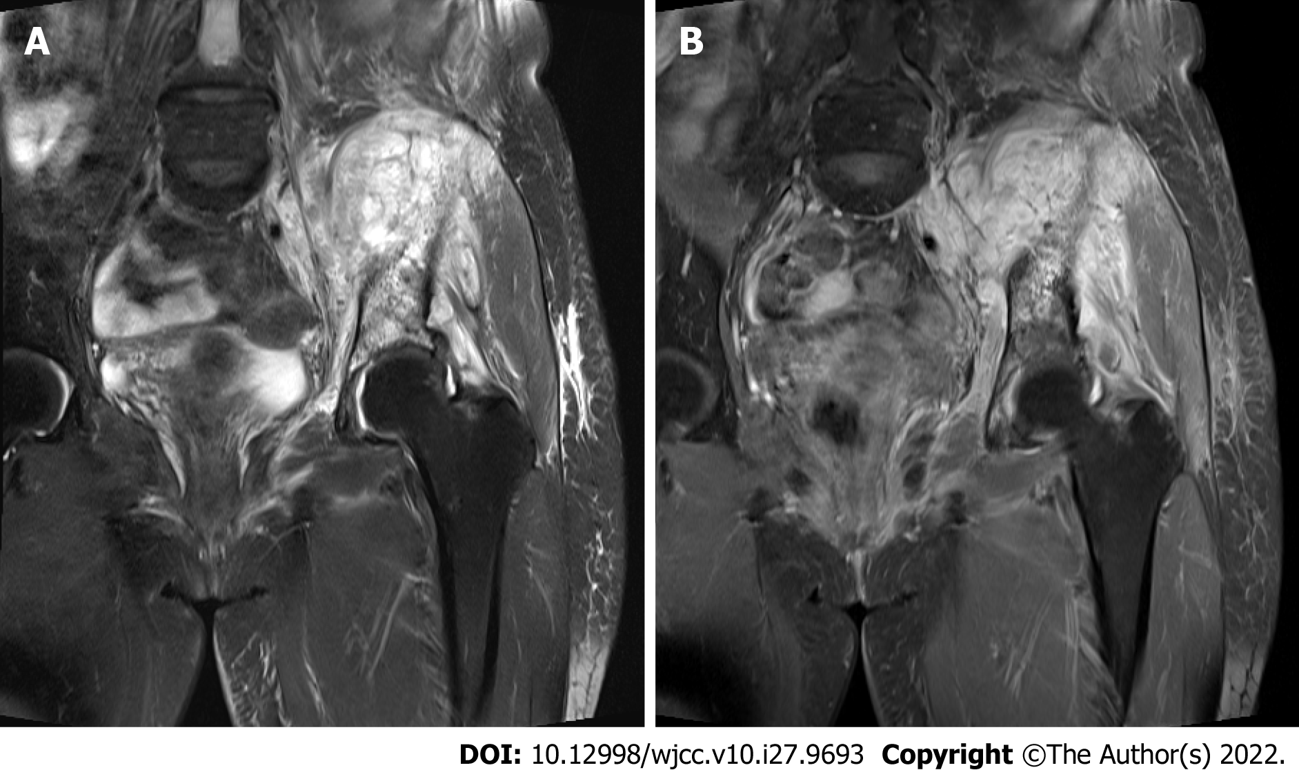
Figure 2 Magnetic resonance image of retroperitoneal sarcoma involved Lt.
pelvis (Lt. iliac bone, Lt. obturator internus muscles, Lt. common and internal iliac lymph node). A: Coronal T2 weighted image; B: Coronal T1 weighted fat suppression image.
- Citation: Song H, Ahn JH, Jung Y, Woo JY, Cha J, Chung YG, Lee KH. Intra and extra pelvic multidisciplinary surgical approach of retroperitoneal sarcoma: Case series report. World J Clin Cases 2022; 10(27): 9693-9702
- URL: https://www.wjgnet.com/2307-8960/full/v10/i27/9693.htm
- DOI: https://dx.doi.org/10.12998/wjcc.v10.i27.9693


