Copyright
©The Author(s) 2022.
World J Clin Cases. Sep 6, 2022; 10(25): 9121-9126
Published online Sep 6, 2022. doi: 10.12998/wjcc.v10.i25.9121
Published online Sep 6, 2022. doi: 10.12998/wjcc.v10.i25.9121
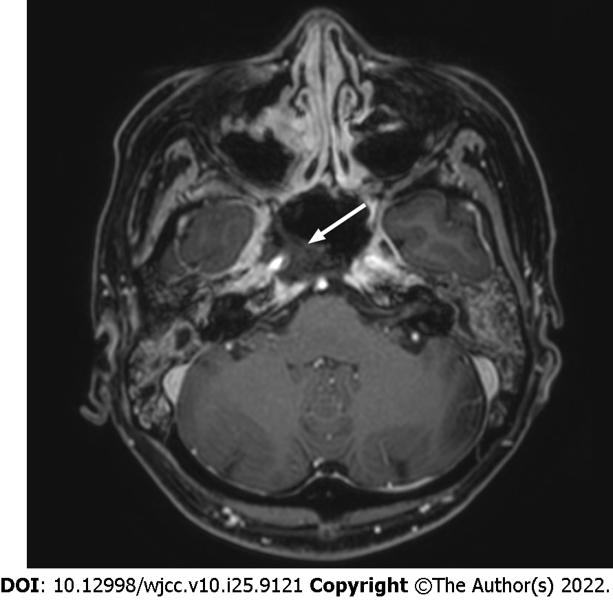
Figure 1 Axial contrast-enhanced T1-weighted magnetic resonance image shows bone destruction in the petrous bone, sphenoid sinus floor, and clivus.
In addition, necrosis of the soft tissues from the nasopharynx to the oropharynx, including the internal carotid artery (white arrow) was observed.
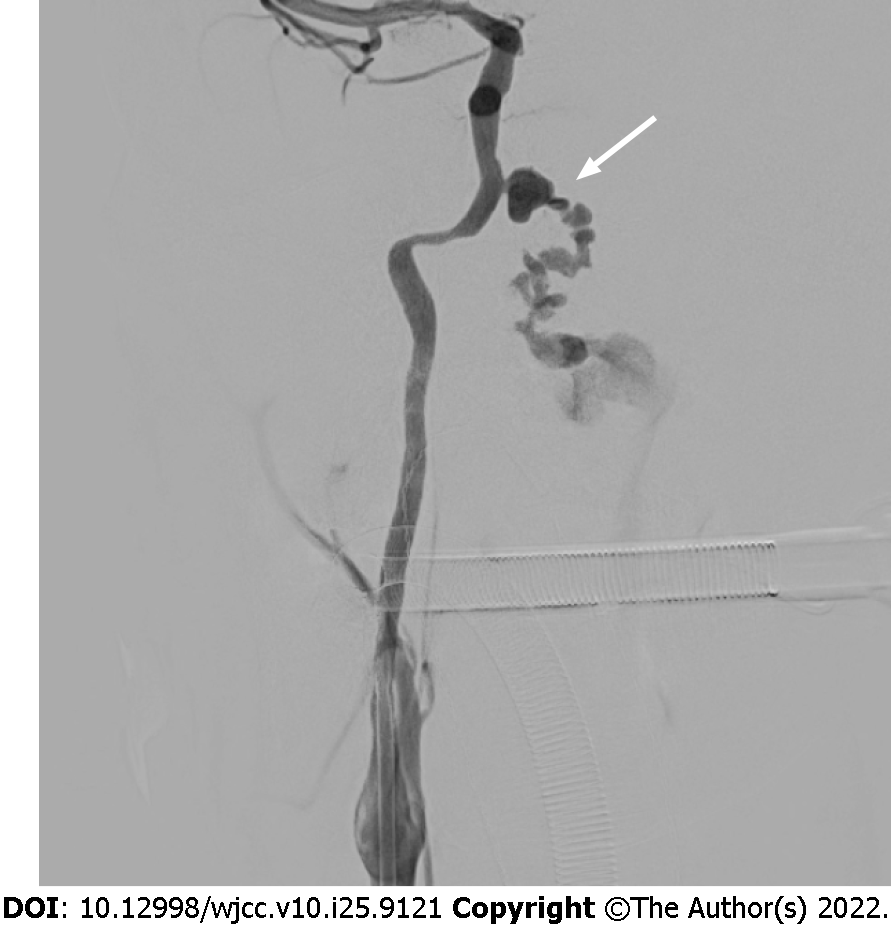
Figure 2
Digital subtraction angiography shows a 9 mm × 5 mm pseudoaneurysm (white arrow) of the right petrous internal carotid artery.
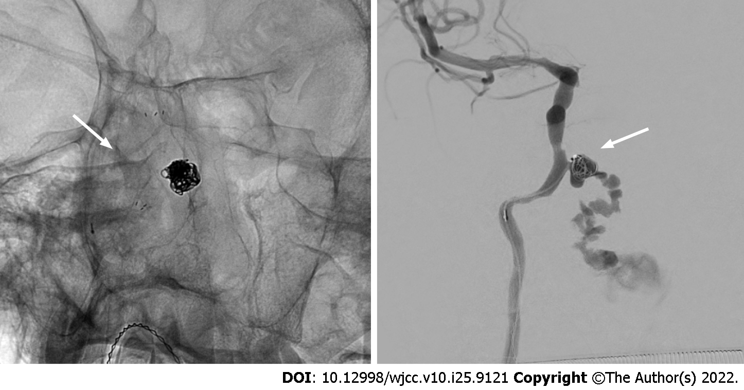
Figure 3 Neuroform Atlas (Boston) 4.
0 mm × 21 mm stent was deployed and then a Guglielmi’s detachable coil was placed into the aneurysm.
Figure 4 Bleeding was observed continuously even after installing the coil into the initially deployed stent located at the orifice of the pseudoaneurysm.
We additionally installed a self-expanding closed-cell low-profile visualized intraluminal support jr 3.5 mm × 18 mm stent.
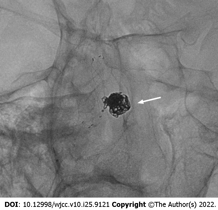
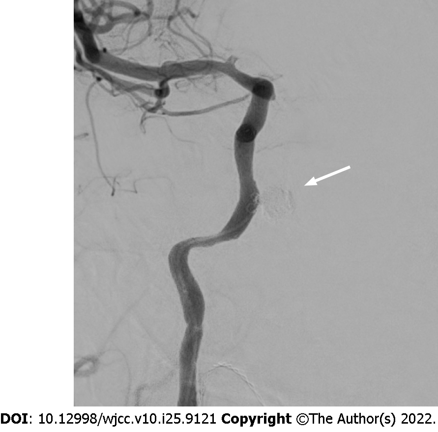
Figure 5 Final angiography reveals complete embolization (white arrow) of pseudoaneurysm.
- Citation: Park JS, Jang HG. Endovascular treatment of a ruptured pseudoaneurysm of the internal carotid artery in a patient with nasopharyngeal cancer: A case report. World J Clin Cases 2022; 10(25): 9121-9126
- URL: https://www.wjgnet.com/2307-8960/full/v10/i25/9121.htm
- DOI: https://dx.doi.org/10.12998/wjcc.v10.i25.9121


