Copyright
©The Author(s) 2022.
World J Clin Cases. Sep 6, 2022; 10(25): 9064-9070
Published online Sep 6, 2022. doi: 10.12998/wjcc.v10.i25.9064
Published online Sep 6, 2022. doi: 10.12998/wjcc.v10.i25.9064
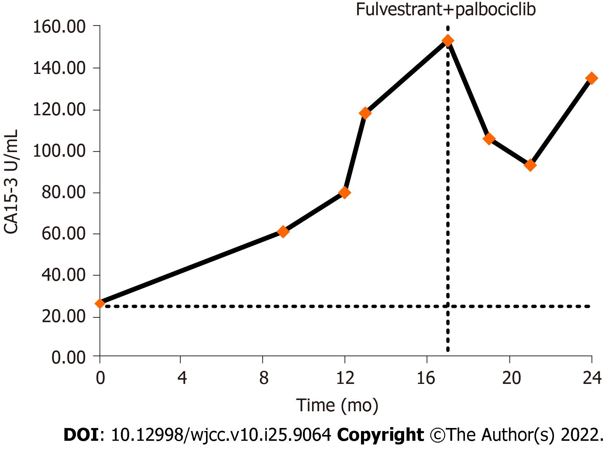
Figure 1 Variation trends of the tumor marker CA15-3.
The X-axis starts at the point when CA15-3 was first detected above the normal range (July 2017) (CA15-3 normal range, 0.0–25.0 U/mL).
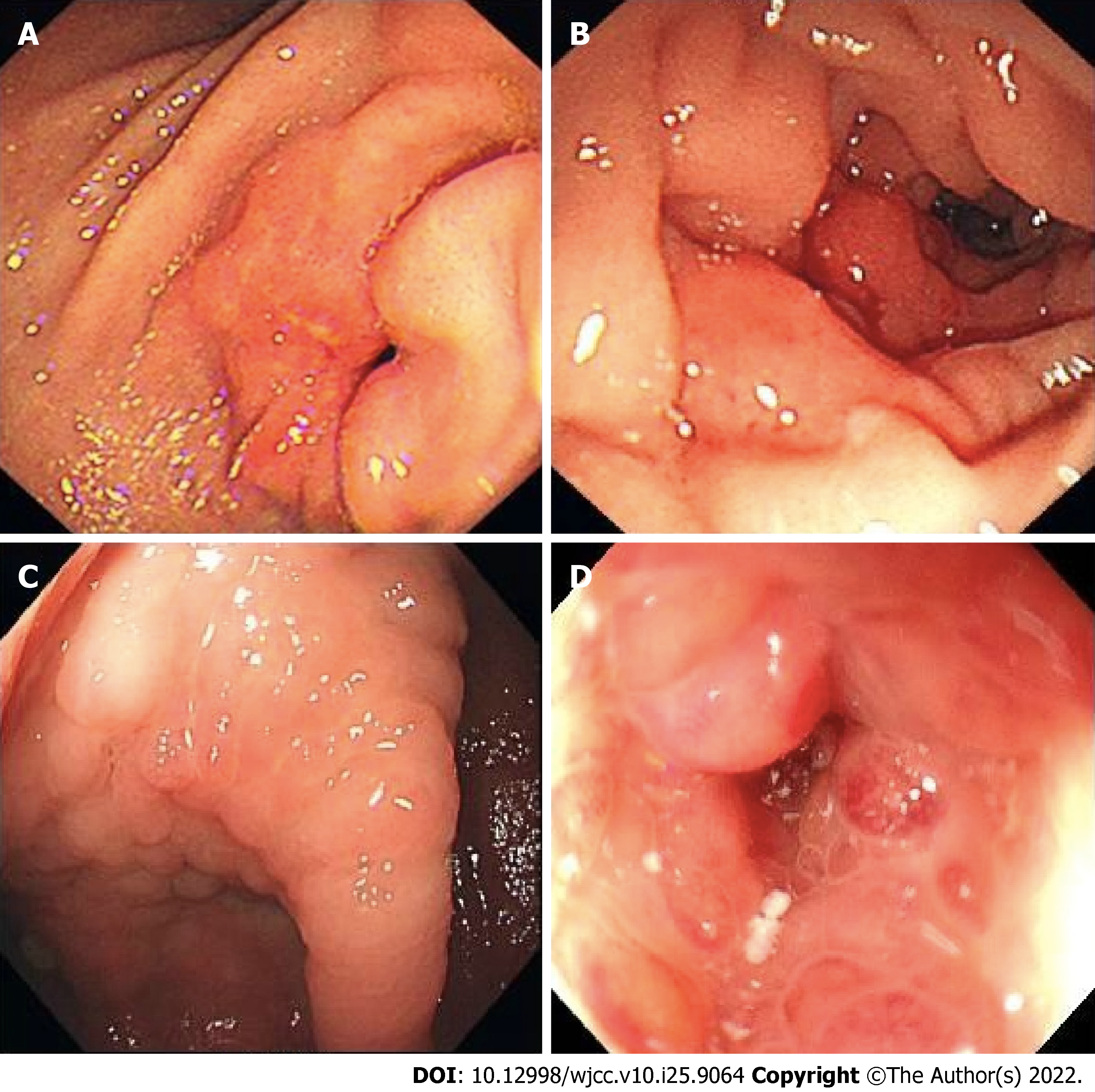
Figure 2 Endoscopic images of gastrointestinal metastasis of breast cancer.
The patient's colonoscopy was performed in another hospital, and the colonoscopy images could not be obtained. A: Endoscopic images of gastric metastasis; B: Duodenal metastasis; C: Ileocecal metastasis; and D: Descending colon metastasis of breast cancer in different patients at our hospital are shown below.
- Citation: Li LX, Zhang D, Ma F. Gastrointestinal metastasis secondary to invasive lobular carcinoma of the breast: A case report. World J Clin Cases 2022; 10(25): 9064-9070
- URL: https://www.wjgnet.com/2307-8960/full/v10/i25/9064.htm
- DOI: https://dx.doi.org/10.12998/wjcc.v10.i25.9064


